|
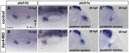
Blockade of her4 has no increase on atoh1b and atoh1a expression. (A–B) Dorsal views of 14 hpf control (A) and her4-MO injected (B) embryos stained by in situ hybridization for atoh1b. atoh1b expression is not affected in the prosensory domains in her4-MO injected embryos. (C–D) Dorsal views of 24 hpf control (C) and her4-MO injected (D) embryos stained by in situ hybridization for atoh1a. atoh1a expression decreases in her4-MO injected embryos. (E–F′) Transversal sections anterior (E, F) and posterior sections (E′, F′) of 28 hpf control (E, E′) and her4-MO injected (F, F′) embryos stained by in situ hybridization for atoh1a. atoh1a expression is as wild-type in her4-MO injected embryos. Dashed circles delineate otic vesicles. All images are at same magnification. Scale bar: 25 µm.
|