
Fig. 2
- ID
- ZDB-FIG-150401-7
- Publication
- Radosevic et al., 2014 - The Role of her4 in Inner Ear Development and Its Relationship with Proneural Genes and Notch Signalling
- Other Figures
- All Figure Page
- Back to All Figure Page
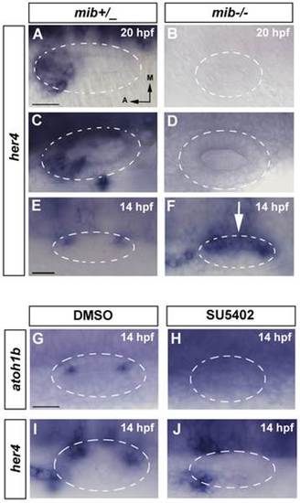
her4 is regulated by Notch in the neurogenic domain but only partially in the sensory domain. (A–F) In situ hybridization for her4 expression in control and mib-/- embryos. (A, C, E) her4 expression in wild-type embryos. At 14 hpf her4 expression is restricted to the two sensory patches (E). At 20 hpf and 24 hpf her4 expression is detected in the sensory and neurogenic domains (A, C). (B, D, F) her4 expression in mib-/- embryos. At 14 hpf her4 expression is detected in the anterior and posterior sensory domains, but it is not repressed in the CMD (white arrow in F). At 20 hpf and 24 hpf its expression in the sensory and neurogenic domains is completely abolished (B, D). (G–J) In situ hybridization for atoh1b and her4 at 14 hpf in DMSO and SU5402 treated embryos. atoh1b (G, H) and her4 expression (I–J) expression is completely lost in embryos treated with SU5402 (H,J) compared with control embryos treated with DMSO (G,I). Dorsal views; anterior to the left, medial to the top. Dashed circles delineate otic vesicles. Images A–D, E–F and G–J are at same magnification. All scale bars: 25 µm. |
| Gene: | |
|---|---|
| Fish: | |
| Condition: | |
| Anatomical Term: | |
| Stage: | 10-13 somites |