
Fig. 3
- ID
- ZDB-FIG-150401-8
- Publication
- Radosevic et al., 2014 - The Role of her4 in Inner Ear Development and Its Relationship with Proneural Genes and Notch Signalling
- Other Figures
- All Figure Page
- Back to All Figure Page
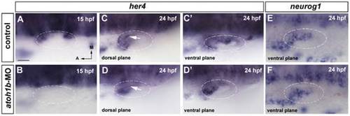
her4 expression requires atoh1b in the sensory domain. (A–D′) In situ hybridization for her4 expression in wild-type and atoh1b-MO injected embryos. (A, B) In 15 hpf control embryos, her4 expression is detected in the future anterior and posterior sensory domains (A), while in morphant embryos her4 expression is lost (B). (C, D) At 24 hpf her4 expression is also lost in the sensory territory in morphant embryos (arrow in D) compared with controls (arrow in C). (C′, D′) The expression in the neurogenic domain is not affected in morphant embryos. (E, F) In situ hybridization for neurog1 expression in wild-type (E) and atoh1b-MO injected (F) embryos. neurog1 expression in atoh1b morphant embryos is not affected. Dorsal views; anterior to the left, medial to the top. Dashed circles delineate otic vesicles. All images are at same magnification. Scale bar: 25 µm. |
| Gene: | |
|---|---|
| Fish: | |
| Knockdown Reagent: | |
| Anatomical Term: | |
| Stage: | Prim-5 |