
- Title
-
Analysis of post-embryonic heart development and maturation in the zebrafish, Danio rerio
- Authors
- Singleman, C., and Holtzman, N.G.
- Source
- Full text @ Dev. Dyn.
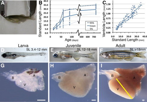
Standard length and cardiac maturation. A: The standard length (SL) of the fish as measured from snout to the base of the tail is a more meaningful measure of maturation state than zebrafish age. B: Zebrafish show a wide range of SL by age. The solid line represents mean growth while upper and lower dashed lines represent 95th and 5th percentiles size range, respectively. C,I: As the zebrafish grows, its cardiac needs grow proportionally, as seen by the linear relationship between SL and ventricle length (VL), measured from the ventricle apex to the bulboventricular junction (C) as indicated with the yellow line in (I). D-F: Maturation of zebrafish from larva (D) to juvenile (E) and finally in adulthood (F) demonstrates the changes in external features. G-I: Cardiac maturation from larva (G) to juvenile (H) and finally in adulthood (I) show not only an increase in heart size, but also changes in cardiac form. BA, bulbous arteriosus; V, ventricle; A, atrium. Scale bar = 100 μm. |
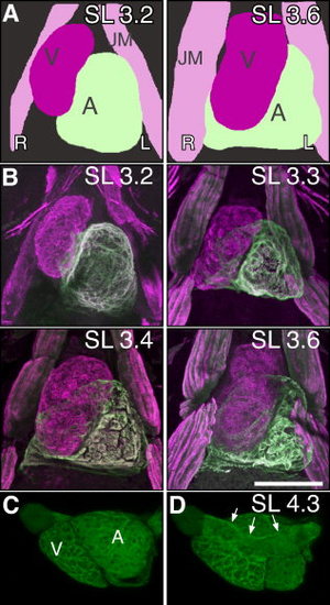
Terminal cardiac rotation. At the transitions between embryonic and larval stages, the atrium moves from the left side of the ventricle to a dorsal location, behind the ventricle. A: Schematic ventral view of embryonic chamber repositioning. The ventricle (magenta) and atrium (light green) are side by side and then the ventricle moves to be more ventral to the atrium. B: Ventral view of muscle marker MF20 antibody (magenta) and the atrial specific marker S46 antibody (green) at standard length (SL) 3.2 (84 hours postfertilization [hpf]) embryo show chambers next to each other. Beginning at SL 3.3 (100 hpf), cardiac rotation initiates. By SL 3.4 (104 hpf), cardiac rotation is complete with the SL 3.6 (120 hpf) heart now in the final orientation. Movies clearly demonstrating the relative three-dimensional organization of the heart at each stage is available as Supp. Movies S1–S4. C, D: While two distinct cardiac chambers are evident in this left lateral view of Tg(myl7:GFP)twu34 labeled heart, the rudiment of the primary heart tube from which the cardiac chambers balloon is visible on the dorsal aspect of the heart (arrows) (D) at SL 4.3. V, ventricle; A, atrium; JM, Jaw Muscles; L, Left; R, Right. Scale bars = 100 μm. EXPRESSION / LABELING:
|
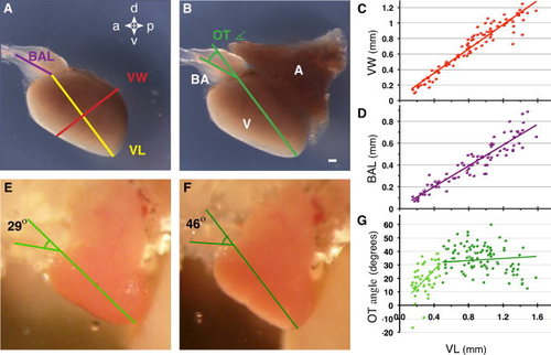
Quantitative cardiac analysis. To better understand cardiac maturation, we carried out a series of quantitative analyses. A,B: Measurements taken for each external cardiac feature; for better characterization of the ventricular morphology, the atrium in (B) has been removed in (A). Bulbous arteriosus length (BAL- purple), ventricle length (VL- yellow), and ventricle width (VW- red) are labeled. B,E,F: The outflow tract angle (OT angle) is indicated in green. C: VW increases linearly with VL. D: BA length increases linearly with VL, staying approximately half the length of the ventricle. The outflow tract (OT) angle changes throughout each cardiac contraction by as much as 30°. E,F: Images extracted from a Supp. Movie S5 demonstrate a minimal OT angle of 29° during ventricular ejection (E) and a maximal OT angle of 46° following atrial systole (F). G: During embryonic and larval development that ends at a VL of 0.5 mm, the OT angle, increases quickly followed by a period of very slow change through adulthood. BA, bulbous arteriosus; V, ventricle; A, atrium; a, anterior; p, posterior; d, dorsal; v, ventral orientation. Scale bar = 100 μm. |
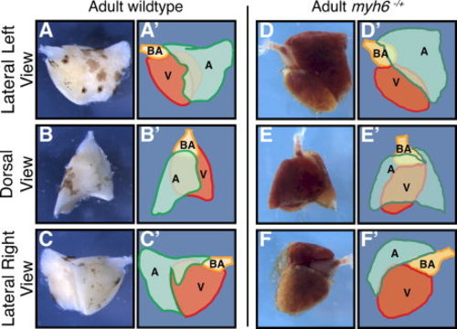
Atrial morphology and its disruption in wea mutants. A–F: The atrium is a U-shaped structure that covers the ventricle, shown here in dorsal, left and right lateral views in wild-type (A–C) and myh6hu423/+ mutants (D–F), paired with a schematic image (A′–C′) and (D′–F′). A,A′: The left side of the ventricle is partially covered by the atrium. B,B′,C,C′: The dorsal aspect of the ventricle is predominantly covered by the atrium while the right side of the ventricle (C,C′) has very limited coverage of the atrium. The atrium in myh6hu423/+ mutants is morphologically distinct from wild-type. D,D′: The left side of the ventricle is partially covered by the atrium and the bulbous arteriosus. E,E′,F,F′ The dorsal aspect of the ventricle is completely covered by the atrium while the right side is barely covered by the atrium (F,F′). A, atrium (green); V, ventricle (red); BA, bulbous arteriosus (orange). PHENOTYPE:
|
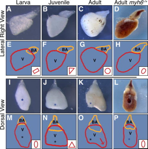
Ventricle morphology maturation and its disruption in wea mutants. Ventricle morphology is observed after removal of the atrium in wild-type (A-C,I-K) and in wea mutants (D,L). A–D: Right lateral images of dissected ventricles. (A) Larval ventricles have a rectangular form (E), (B) juvenile heart are triangular (F), while (C) adult heart have a round-shaped ventricle (G). E–H: Tracings of the ventricle (V, red) and bulbous arteriosus (BA, orange) with representative ventricle morphology in the lower right-hand corner. D,H: The ventricle in wea mutant adults (D) fails to mature and appears oval (H). I-L: Dorsal images of the same ventricles as in A-D. I,M: From a dorsal view larval heart (I) have an oval form (M). (J) juvenile hearts have a triangular shape (N) and (K) adult hearts have an extension (arrow) of the ventricle opposite of the atrioventricular valve (O). L,P: The immature oval morphology of the wea ventricle is also evident when viewed dorsally. M–P: Tracings of the ventricle and bulbous with standard shapes in the lower right-hand corner. The (*) indicates the atrioventricular valve location. PHENOTYPE:
|
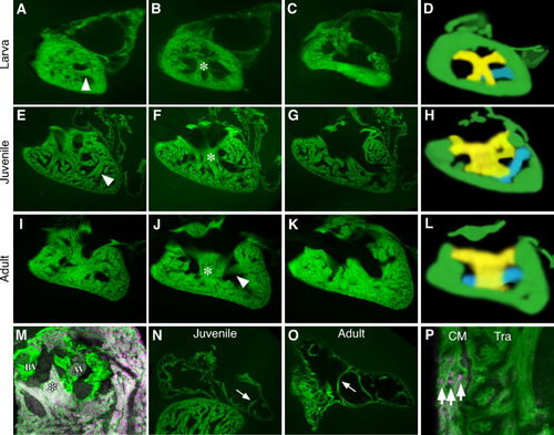
Internal chamber organization. A-L: The ventricular myocardium is highly trabeculated by the early larval stages and becomes progressively more organized during maturation. A-L: Serial sections of Tg(myl7:GFP)twu34 hearts during different stages of maturation. A–C are larval, E-G are juvenile, and I–K are representative adult hearts. During maturation, two clear muscle groups become apparent. A transverse band (arrowhead) can be seen in A, E, and J and is depicted in blue on the diagram in D, H and L. A tri-tipped muscle (*) in B, F and J and depicted in yellow on the diagram in D, H and L. (M) The tri-tipped muscle (false-colored magenta with Tg(-5.1myl7:nDsRed2)f2) can be seen attached the base of the AV and BV valves (labeled green with Tg(fli1a:EGFP)y1). The atrium is significantly less trabeculated than the ventricle even during larval and adult stages however pectinate muscles are observed (arrows in N,O). P demonstrates the presence of a clear ventricular compact myocardium (CM) adjacent to the trabeculae (Tra) that is three cell layers thick in this cross section of an adult heart (arrows) with myocardial nuclei false-colored magenta using Tg(-5.1myl7:nDsRed2)f2 and the myocardial cells green with Tg(myl7:GFP)twu34. EXPRESSION / LABELING:
|

Unillustrated author statements PHENOTYPE:
|