|
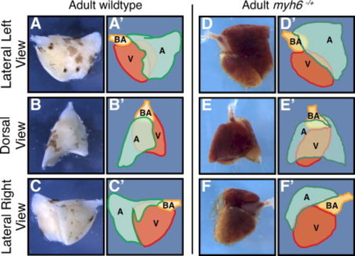
Atrial morphology and its disruption in wea mutants. A–F: The atrium is a U-shaped structure that covers the ventricle, shown here in dorsal, left and right lateral views in wild-type (A–C) and myh6hu423/+ mutants (D–F), paired with a schematic image (A′–C′) and (D′–F′). A,A′: The left side of the ventricle is partially covered by the atrium. B,B′,C,C′: The dorsal aspect of the ventricle is predominantly covered by the atrium while the right side of the ventricle (C,C′) has very limited coverage of the atrium. The atrium in myh6hu423/+ mutants is morphologically distinct from wild-type. D,D′: The left side of the ventricle is partially covered by the atrium and the bulbous arteriosus. E,E′,F,F′ The dorsal aspect of the ventricle is completely covered by the atrium while the right side is barely covered by the atrium (F,F′). A, atrium (green); V, ventricle (red); BA, bulbous arteriosus (orange).
|