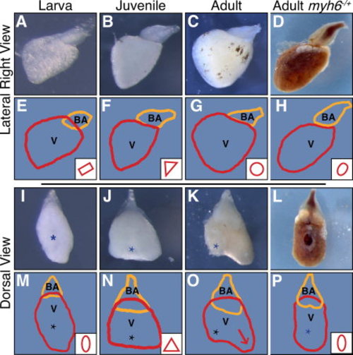
Ventricle morphology maturation and its disruption in wea mutants. Ventricle morphology is observed after removal of the atrium in wild-type (A-C,I-K) and in wea mutants (D,L). A–D: Right lateral images of dissected ventricles. (A) Larval ventricles have a rectangular form (E), (B) juvenile heart are triangular (F), while (C) adult heart have a round-shaped ventricle (G). E–H: Tracings of the ventricle (V, red) and bulbous arteriosus (BA, orange) with representative ventricle morphology in the lower right-hand corner. D,H: The ventricle in wea mutant adults (D) fails to mature and appears oval (H). I-L: Dorsal images of the same ventricles as in A-D. I,M: From a dorsal view larval heart (I) have an oval form (M). (J) juvenile hearts have a triangular shape (N) and (K) adult hearts have an extension (arrow) of the ventricle opposite of the atrioventricular valve (O). L,P: The immature oval morphology of the wea ventricle is also evident when viewed dorsally. M–P: Tracings of the ventricle and bulbous with standard shapes in the lower right-hand corner. The (*) indicates the atrioventricular valve location.
|
