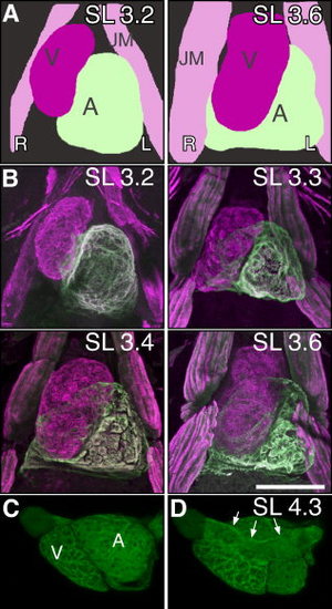
Terminal cardiac rotation. At the transitions between embryonic and larval stages, the atrium moves from the left side of the ventricle to a dorsal location, behind the ventricle. A: Schematic ventral view of embryonic chamber repositioning. The ventricle (magenta) and atrium (light green) are side by side and then the ventricle moves to be more ventral to the atrium. B: Ventral view of muscle marker MF20 antibody (magenta) and the atrial specific marker S46 antibody (green) at standard length (SL) 3.2 (84 hours postfertilization [hpf]) embryo show chambers next to each other. Beginning at SL 3.3 (100 hpf), cardiac rotation initiates. By SL 3.4 (104 hpf), cardiac rotation is complete with the SL 3.6 (120 hpf) heart now in the final orientation. Movies clearly demonstrating the relative three-dimensional organization of the heart at each stage is available as Supp. Movies S1–S4. C, D: While two distinct cardiac chambers are evident in this left lateral view of Tg(myl7:GFP)twu34 labeled heart, the rudiment of the primary heart tube from which the cardiac chambers balloon is visible on the dorsal aspect of the heart (arrows) (D) at SL 4.3. V, ventricle; A, atrium; JM, Jaw Muscles; L, Left; R, Right. Scale bars = 100 μm.
|
