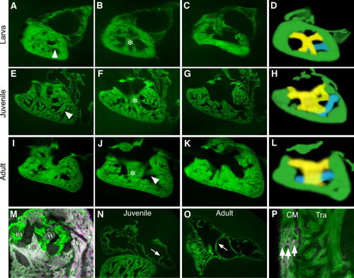
Internal chamber organization. A-L: The ventricular myocardium is highly trabeculated by the early larval stages and becomes progressively more organized during maturation. A-L: Serial sections of Tg(myl7:GFP)twu34 hearts during different stages of maturation. A–C are larval, E-G are juvenile, and I–K are representative adult hearts. During maturation, two clear muscle groups become apparent. A transverse band (arrowhead) can be seen in A, E, and J and is depicted in blue on the diagram in D, H and L. A tri-tipped muscle (*) in B, F and J and depicted in yellow on the diagram in D, H and L. (M) The tri-tipped muscle (false-colored magenta with Tg(-5.1myl7:nDsRed2)f2) can be seen attached the base of the AV and BV valves (labeled green with Tg(fli1a:EGFP)y1). The atrium is significantly less trabeculated than the ventricle even during larval and adult stages however pectinate muscles are observed (arrows in N,O). P demonstrates the presence of a clear ventricular compact myocardium (CM) adjacent to the trabeculae (Tra) that is three cell layers thick in this cross section of an adult heart (arrows) with myocardial nuclei false-colored magenta using Tg(-5.1myl7:nDsRed2)f2 and the myocardial cells green with Tg(myl7:GFP)twu34.
|
