
- Title
-
Phylogeny of the teashirt-related zinc finger (tshz) gene family and analysis of the developmental expression of tshz2 and tshz3b in the zebrafish
- Authors
- Santos, J.S., Fonseca, N.A., Vieira, C.P., Vieira, J., and Casares, F.
- Source
- Full text @ Dev. Dyn.
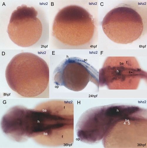
Spatial and temporal expression pattern of tshz2 detected by RNA in situ hybridization. A-D: Embryos at 2 (A), 4 (B), 6 (C), and 8 (D) hours postfertilization (hpf). tshz2 mRNA is detected in early stages but signal fades by 8-10 hpf. Re-expression of tshz2 occurs around the prim-5 stage (24 hpf) (E,F). G,H: tshz2 expression pattern in a 36 hpf embryo. At 36 hpf, tshz2 is also expressed in the liver primordium (not shown). F,G: Dorsal views with anterior to the left. (E and H) Lateral views with dorsal up and anterior to the left. ba, branchial arches; f, pectoral fin; h, hindbrain; l, liver; op, olfactory placode; sc, spinal cord. |
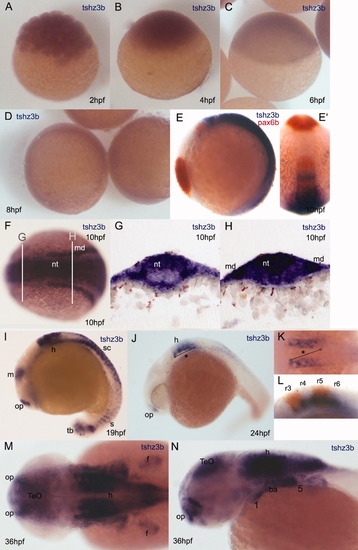
Spatial and temporal expression pattern of tshz3b detected by RNA in situ hybridization. A-D: Embryos at 2 (A), 4 (B), 6 (C), and 8 (D) hours postfertilization (hpf). tshz3b mRNA is detected in early stages but signal fades by 8 hpf. E,F: At 10 hpf, tshz3b expression resumes and is present from intermediate until the caudal region of the neural tube: lateral (E) and dorsal (F) views. The embryo in E is costained with a pax6b antisense RNA probe. (brown). (E′ is a dorsal view of the same embryo; rostral is up). G,H: Transversal Vibratome sections of a 10 hpf embryo at the anteroposterior levels marked in F. In G, tshz3b is expressed in the dorsal neural tube (nt), but in its most posterior regions (H), tshz3b expression is present both dorsally and ventrally. (md) in (H) marks tshz3b expression in the mesoderm (md). I: At the 20-somite stage (19 hpf), tshz3b is expressed in the olfactory placodes, midbrain, hindbrain, spinal cord, somites, and tail bud. J: At prim-5 stage (24 hpf), tshz3b expression is present in the olfactory placodes and in the hindbrain. K: Dorsal view at the hindbrain level (*) of the embryo shown in J. L: Lateral view of the hindbrain of a 24 hpf embryo marked for tshz3b (blue) and krox20 (brown); tshz3b is expressed from r3 to r6. M,N: tshz3b expression in the anterior region of a 36 hpf embryo. Dorsal (M) and lateral (N) views are shown. ba, branchial arches; f, pectoral fin; h, hindbrain; m, midbrain; md, mesoderm; nt, neural tube; op, olfactory placode; r3-6, rhombomeres 3 to 6; s, somites; sc, spinal cord; tb, tail bud; TeO, tectum opticum. |
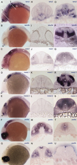
Spatial expression pattern of tshz2, tshz3b, meis1, meis2.1, meis2.2, pax6a, and pax6b in 24 hours postfertilization (hpf) embryos revealed by RNA in situ hybridization. A-G: Lateral views with dorsal up and anterior to the left. H-U: Transversal vibratome sections at the eyes (left pictures) and rhombomere 5 (right pictures) at the level marked by the lines (1 and 2, respectively) in A. A-G: All genes are expressed in the hindbrain. In Vibratome sections (O-U), it is possible to observe that tshz2 coexpresses with tshz3b, meis, and pax6a, although at different dorsoventral levels. tshz and meis genes also coexpress in the olfactory placodes (A-E) and tshz2 and all the meis genes coexpress in the spinal cord (A,C,D,E). Lines (1) and (2) in A mark the approximate section planes shown in the middle and right columns. ba, branchial arches; d, diencephalon; h, hindbrain; m, midbrain; md, mesoderm; nt, neural tube; op, olfactory placode; r, retina; sc, spinal cord. EXPRESSION / LABELING:
|
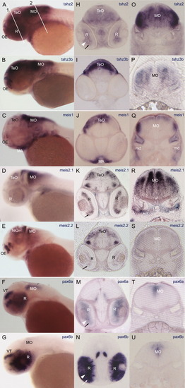
Spatial expression pattern of tshz2, tshz3b, meis1, meis2.1, meis2.2, pax6a, and pax6b in 48 hours postfertilization (hpf) embryos revealed by RNA in situ hybridization. A-G: Lateral views with dorsal up and anterior to the left. H-U: Transversal vibratome sections at the eyes (left pictures) and rhombomere5 (right pictures) level. A-G,O-U: All genes are expressed in medulla oblongata. H-L: tshz2 is expressed in the midbrain (H) such as tshz3b and meis genes, although coexpression takes place at different dorsoventral levels (H-L). A,B,C,E: tshz2 coexpresses with tshz3b, meis1, and meis2.2 in the olfactory epithelium. In H,K,L,M,N, it is possible to observe that tshz2, meis2.1, meis2.2, pax6a, and pax6b, respectively, are expressed in the neural retina. Lines (1) and (2) in (A) mark the approximate section planes shown in the middle and right columns. MA, mandibular arch; md, mesoderm; MO, medulla oblongata; OE, olfactory epithelium; R, retina; TeO, tectum opticum; Th, thalamus; PTv, ventral posterior tuberculum; VT, ventral tectum. Arrowhead: ganglion cell layer. Arrow: Inner nuclear layer. EXPRESSION / LABELING:
|
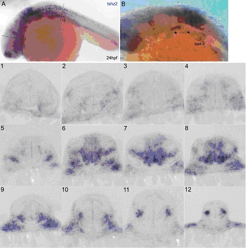
tshz2 is expressed along the hindbrain and spinal cord and in branchial arches 4-5 in 24hpf embryos EXPRESSION / LABELING:
|
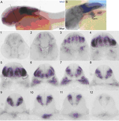
tshz2 is expressed along the hindbrain and spinal cord and in branchial arches 4-5 of 36hpf embryos. EXPRESSION / LABELING:
|
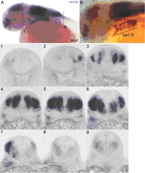
tshz3b is expressed along the hindbrain, in the anterior spinal cord and in branchial arches 1-5 of 36hpf embryos. EXPRESSION / LABELING:
|

Unillustrated author statements |