
|
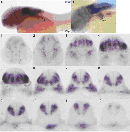
Fig. S3
tshz2 is expressed along the hindbrain and spinal cord and in branchial arches 4-5 of 36hpf embryos.
In situ hybridization of 36hpf embryos using a tshz2 RNA probe. (A) Lateral view with dorsal up and anterior to the left. (1-12) 40μm transversal vibratome sections of the embryo in (A) at different AP levels. First and last sections are represented in (A) as 1 and 12, respectively. (1, 2) Faint expression is detected in the midbrain and becomes stronger in the hindbrain (3-12). Here, around the otic vesicle level (4,5), tshz2 is expressed in medial and lateral regions of the medulla oblongata in the ventral neural tube. More posteriorly (7-12), the expression becomes restricted to the lateral portion of the medulla oblongata. The expression in the lateral cell cluster along the spinal cord that was detected at 24hpf is no longer seen. (B) Dorso-lateral view with dorsal up and anterior to the left at the branchial arches (ba) level. tshz2 is expressed in pharyngeal arches 4 and 5.