
Fig. 5
|
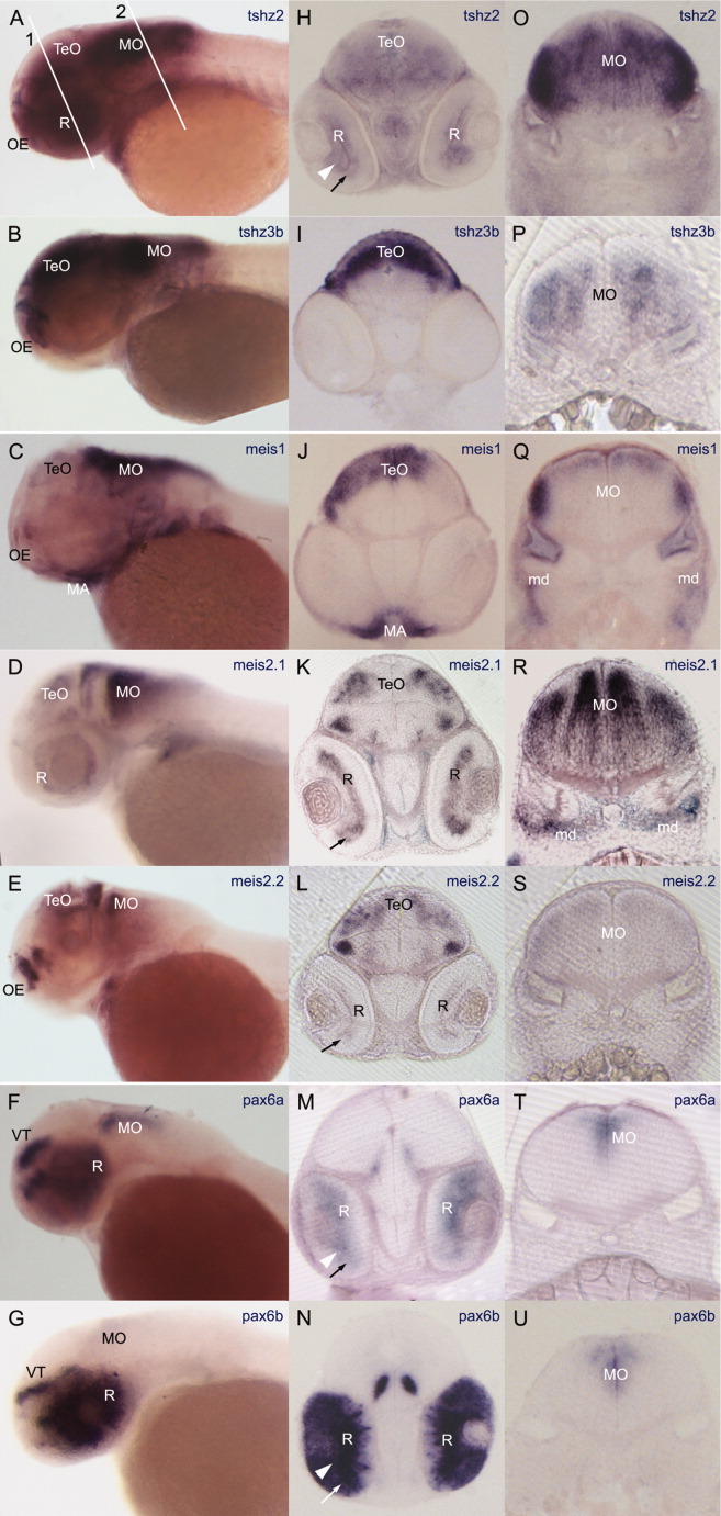
Fig. 5 Spatial expression pattern of tshz2, tshz3b, meis1, meis2.1, meis2.2, pax6a, and pax6b in 48 hours postfertilization (hpf) embryos revealed by RNA in situ hybridization. A-G: Lateral views with dorsal up and anterior to the left. H-U: Transversal vibratome sections at the eyes (left pictures) and rhombomere5 (right pictures) level. A-G,O-U: All genes are expressed in medulla oblongata. H-L: tshz2 is expressed in the midbrain (H) such as tshz3b and meis genes, although coexpression takes place at different dorsoventral levels (H-L). A,B,C,E: tshz2 coexpresses with tshz3b, meis1, and meis2.2 in the olfactory epithelium. In H,K,L,M,N, it is possible to observe that tshz2, meis2.1, meis2.2, pax6a, and pax6b, respectively, are expressed in the neural retina. Lines (1) and (2) in (A) mark the approximate section planes shown in the middle and right columns. MA, mandibular arch; md, mesoderm; MO, medulla oblongata; OE, olfactory epithelium; R, retina; TeO, tectum opticum; Th, thalamus; PTv, ventral posterior tuberculum; VT, ventral tectum. Arrowhead: ganglion cell layer. Arrow: Inner nuclear layer.