
|
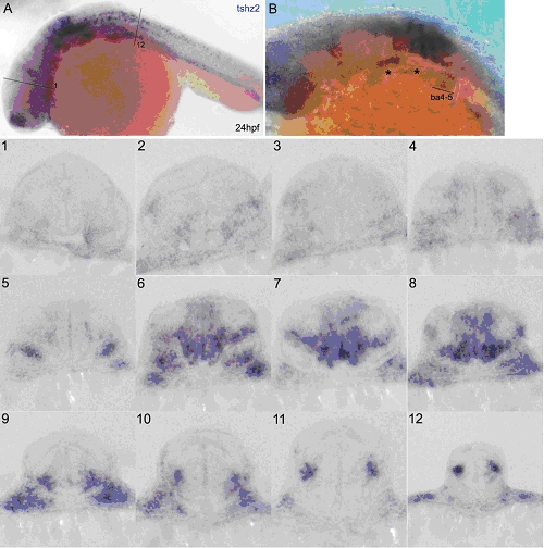
Fig. S2
tshz2 is expressed along the hindbrain and spinal cord and in branchial arches 4-5 in 24hpf embryos
In situ hybridization of 24hpf embryos using a tshz2 RNA probe. (A) Lateral view with dorsal up and anterior to the left. (B) Dorso-lateral view with dorsal up and anterior to the left focused at the branchial arches (ba) level. (1-12) 40µm transversal vibratome sections of the embryo in (A) at different anteriorposterior (AP) levels. First and last sections are represented in as 1 and 12, respectively. tshz2 expression in the hindbrain varies along the AP axis: its expression is stronger in rhombomeres 4 to 6 (sections no. 6-8). Along the dorsal-ventral extent, tshz2 is expressed more strongly in ventro-medial and ventro-lateral portions of the hindbrain. At spinal cord level, tshz2 expression is restricted to a lateral cell cluster (pictures 10-12). (B) tshz2 is expressed in branchial arches 4 and 5. Some signal is detected in more anterior branchial arches (asterisks).