
- Title
-
Cystein-serine-rich nuclear protein 1, Axud1/Csrnp1, is essential for cephalic neural progenitor proliferation and survival in zebrafish
- Authors
- Feijoo, C.G., Sarrazin, A.F., Allende, M.L., and Glavic, A.
- Source
- Full text @ Dev. Dyn.
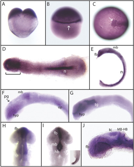
Expression of zebrafish axud1 gene. Detection of axud1 mRNA was carried out by whole-mount in situ hybridization using different stage embryos from two cells to 24 hpf. Images in A, B, E, F, G, and J are lateral views, C is a dorso-anterior view, and D, H, and I are dorsal views. A: Two-cell-stage embryo. B: Thirty percent epiboly. axud1 is detected ubiquitously with higher expression at the marginal zone (arrow) and animal pole. C: A 10-hpf embryo. Expression is detected in the prechordal plate and chordamesoderm, the anterior limit is indicated (arrow). D: A 10-hpf flat-mount embryo. axud1 expression localises in the head and polster regions (bracket) and notochord (n). E: A 12-hpf embryo. Expression is observed in the anterior forebrain, midbrain, and notochord. F: A 16-hpf embryo. axud1 mRNA is found in the anterior hypothalamus, pineal gland (arrow), dorsal midbrain region, and scattered cells in the otic placode. At 20 hpf (G, H) and 22 hpf (I), the expression becomes restricted to the forebrain and midbrain brain ventricles and the tip of the tail (inset in I). J: A 24-hpf embryo. axud1 RNA appears in cephalic regions with higher levels at the forebrain, tectum, midbrain-hindbrain boundary, and hindbrain. fb, forebrain; mb, midbrain; hb, hindbrain; n, notochord; tc, tectum; tg, tegmentum; pg, pineal gland; hyp, hypothalamus; MB-HB, midbrain-hindbrain boundary; op, otic placode. |

axud1 morphant embryos show defects in head size but not in A/P patterning or differentiation. A-D: Wild type embryos. E-H: axud1 morphant embryos. A, E: Images of live wild type (A) and morphant embryos (E). The arrow indicates the eyes and the discontinued line encircles the otic vesicle. Note the evident reduction in eye size. B, F: Expression patterns of the forebrain marker emx, the midbrain marker pax2.1, and the hindbrain marker krox20 in wild type (B) and morphant embryos (F). No variations in the antero-posterior brain pattern are observed. C, D, G, H: Immuno stain for the neuronal markers acetylated tubulin (C, G) and islet-1 (D, H) in wild type and morphant embryos. Although the brain region is reduced in size, neural differentiation as well as neuronal connectivity appears normal. MB-HB, midbrain-hindbrain boundary; r3, rhombomere 3; r5, rhombomere 5. tel, telencephalon; tc, tectum; tg, trigeminal ganglion. All panels show lateral views of 24-hpf embryos; dorsal is up, anterior is left. |
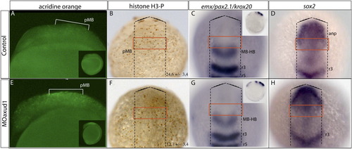
axud1 morphant embryos have decreased proliferation and extensive cell death in the presumptive midbrain region. A-D: Wild type 10-hpf embryos. E-H: axud1 morphant embryos. A, E: Cell death analysis revealed by acridine orange staining of wild type (A) and morphant embryos (E). Cell death is mainly detected in the presumptive midbrain region (pMB). B, F: Proliferation analysis using the anti-phospho-histone H3 (H3-P) immunostain in wild type (B) and morphant embryos (F). The discontinuous line demarcates the embryo. The number of H-P3-positive cells quantified within the red square is markedly reduced in morphant embryos compared with wild type (24.6 ± 3.4 vs. 12.7 ± 3.4, n = 15 embryos; P < 0.001). C, G: Expression pattern of the forebrain marker emx, the midbrain marker pax2.1, and the hindbrain marker krox20 in wild type (C) and morphant embryos (G). No significant variations in the antero-posterior brain pattern are observed at this early stage. D, H: Expression pattern of the neural progenitor marker sox2 in wild type (D) and morphant embryos (H). No alterations in neural progenitor distribution or intensity of the signal are recognized. anp, anterior neural plate; MB-HB, midbrain-hindbrain boundary; pMB, presumptive midbrain; r3, rhombomere 3; r5, rhombomere 5. Lateral (A, B, E, F) and dorsal (C, D, G, H) views of 10-hpf embryos. |

Axud1 gain of function rescues the morphant phenotype. A-D: Live embryos. E-H: Acridine orange stain of the same embryos that are depicted in A-D. Insets show magnifications of the region encompassed by the brackets. A, E: Control embryos; no cell death is detected (E). B, F: Morphant embryos. Notice the drastic decrease in the eye size (B) and the extensive cell death in the midbrain region (F, bracket and inset). C, G: Splice morpholino and axud1 mRNA co-injection significantly rescue the head and eye size reductions and the cell death phenotype. D, H: The overexpression of Axud1 has no obvious phenotype. The phenotypes depicted in each panel correspond to representative embryos and the corresponding percentages are: 80% showing this phenotype in morphant embryos (n = 93), 60% of embryos show rescue (n = 87), and 100% of mRNA injected embryos appear normal (n = 96). I: RT-PCR showing the efficiency of the splice morpholino to block correct splicing of the axud1 transcript. cDNA was prepared from uninjected (“C”), co-injected with morpholino, and axud1 mRNA (“MO+mRNA”), or injected with axud1 mRNA (“mRNA”) embryos. The panel shows the internal control (β-actin, upper band) and the spliced axud1 mRNA (lower band). The absence of the axud1 band in MO-injected embryos (“MO”) indicates defective axud1 pre-RNA splicing. Note the strong axud1 band in embryos injected with the axud1 mRNA showing the presence of exogenous message. The bracket indicates the midbrain region and the dashed line indicate the eye. MB-HB, midbrain-hindbrain boundary. All panels are lateral views. PHENOTYPE:
|
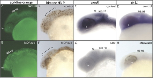
Shh, Six3.1, and Axud1 interactions. A-D: Wild type embryos. E-H: axud1 morphant embryos. A, E: Acridine orange stain of control (A) and experimental embryos (E). Cell death is detected throughout the cephalic region. B, F: Proliferation analysis evidenced by the anti-phospho-histone H3 (H3-P) immunostain in wild type (B) and axud1 morphants (F). Note the reduced number of H3-P-labelled cells along the bracket in morphant embryos compared with the wild type siblings. C, G: axud1 in situ hybridization in control (C) and in smub577 mutant embryos (G). An evident decrease in axud1 expression is detected in embryos with reduced levels of Shh pathway activity. Inset in G shows that in situ stain has labeled the notochord but not the head region. D, H: six3.1 in situ hybridization in wild type (D) and axud1 morphant embryos (I). Strong inhibition of six3.1 transcription is observed in experimental embryos. MB-HB, midbrain-hindbrain boundary; e, eye field; t, telencephalon; tc, tectum. All panels are lateral views of 24-hpf embryos. |
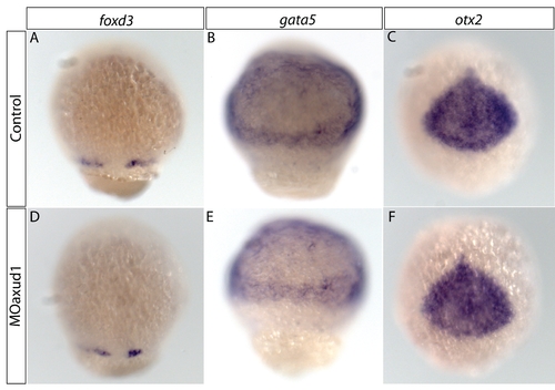
Axud1 does not alter specification of the germ layers. (A-C) Wild type embryos and (D-F) axud1 morphant embryos at 70% epiboly. In situ hibridization analysis of foxd3 expression in the mesoderm (A, D); gata5 expression in mesendoderm (B, E), and otx2 in anterior neural ectoderm (C, F). The normal expression of these markers indicates that there is no alteration in the specification in any of the three germ layers in morphant embryos. |
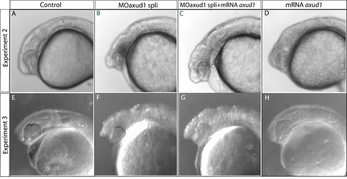
Axud1 gain of function rescues the morphant phenotype: additional examples. A-D: Live embryos. A, E: Control embryos. B, F: axud1 splice morphant embryos. C, G: Splice morpholino and axud1 mRNA co-injected embryos. D, H: axud1 mRNA overexpression embryos. Pictures show representative embryos from two additional independent rescue experiments. All panels are lateral views and anterior is to the left. |