
|
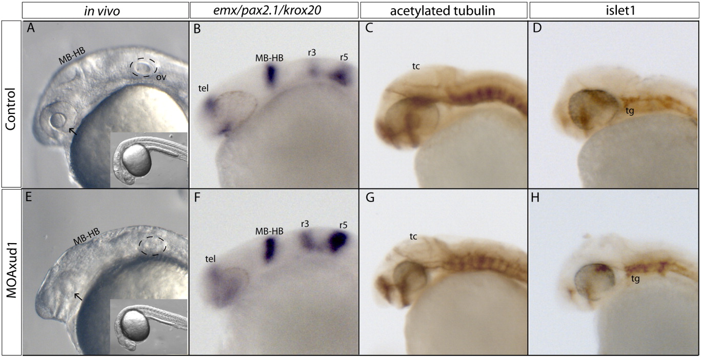
Fig. 3 axud1 morphant embryos show defects in head size but not in A/P patterning or differentiation. A-D: Wild type embryos. E-H: axud1 morphant embryos. A, E: Images of live wild type (A) and morphant embryos (E). The arrow indicates the eyes and the discontinued line encircles the otic vesicle. Note the evident reduction in eye size. B, F: Expression patterns of the forebrain marker emx, the midbrain marker pax2.1, and the hindbrain marker krox20 in wild type (B) and morphant embryos (F). No variations in the antero-posterior brain pattern are observed. C, D, G, H: Immuno stain for the neuronal markers acetylated tubulin (C, G) and islet-1 (D, H) in wild type and morphant embryos. Although the brain region is reduced in size, neural differentiation as well as neuronal connectivity appears normal. MB-HB, midbrain-hindbrain boundary; r3, rhombomere 3; r5, rhombomere 5. tel, telencephalon; tc, tectum; tg, trigeminal ganglion. All panels show lateral views of 24-hpf embryos; dorsal is up, anterior is left.