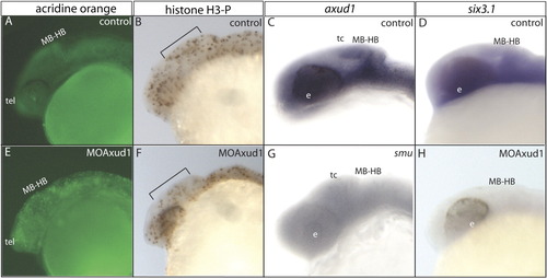
Shh, Six3.1, and Axud1 interactions. A-D: Wild type embryos. E-H: axud1 morphant embryos. A, E: Acridine orange stain of control (A) and experimental embryos (E). Cell death is detected throughout the cephalic region. B, F: Proliferation analysis evidenced by the anti-phospho-histone H3 (H3-P) immunostain in wild type (B) and axud1 morphants (F). Note the reduced number of H3-P-labelled cells along the bracket in morphant embryos compared with the wild type siblings. C, G: axud1 in situ hybridization in control (C) and in smub577 mutant embryos (G). An evident decrease in axud1 expression is detected in embryos with reduced levels of Shh pathway activity. Inset in G shows that in situ stain has labeled the notochord but not the head region. D, H: six3.1 in situ hybridization in wild type (D) and axud1 morphant embryos (I). Strong inhibition of six3.1 transcription is observed in experimental embryos. MB-HB, midbrain-hindbrain boundary; e, eye field; t, telencephalon; tc, tectum. All panels are lateral views of 24-hpf embryos.
|
