
- Title
-
Mutations affecting somite formation and patterning in the zebrafish, Danio rerio
- Authors
- van Eeden, F.J., Granato, M., Schach, U., Brand, M., Furutani-Seiki-, M., Haffter, P., Hammerschmidt, M., Heisenberg, C.P., Jiang, Y.J., Kane, D.A., Kelsh, R.N., Mullins, M.C., Odenthal, J., Warga, R.M., Allende, M.L., Weinberg, E.S., and Nüsslein-Volhard, C.
- Source
- Full text @ Development
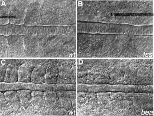
Dorsal view of live embryos mutant for fss (B), bea (D) and their wild-type siblings (A and C, respectively). In A the sixth somite is just forming. (B) fss mutant at the same A/P level; no somite boundaries are visible. In C the seventh somite is just forming at the left of the picture. (D) bea mutant at the same level as C. Boundaries are present, but indistinct and irregularly placed. PHENOTYPE:
|
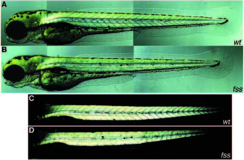
An overview of the fss phenotype at 84 hpf. (A) Wild-type sibling, (B) homozygous mutant; no V-shaped myotomes are present. Usually mutants are slightly shorter. (C,D) Same embryos, dark-field image. Striated muscle shows a bright birefringence. Segment borders are visible as dark lines. PHENOTYPE:
|
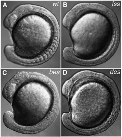
Comparison of the three phenotypic groups of somite boundary mutants. All embryos are at the 11- to 13-somite stage. (A) Wild-type embryo. (B) fss mutant; only very few boundaries are present anteriorly. (C) bea mutant; in this individual the phenotype seems almost as severe as in fss; normally the anterior somites are more regular. (D) des mutant; the first eight somites look normal; no somites are formed beyond this point. The somite phenotype of aei and wit is similar to that of des. PHENOTYPE:
|
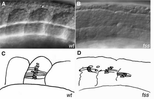
Adaxial cells differentiate normally in fss mutants. (A) Somite 7 at the 13-somite stage, lateral view. At the middle of the somite a stack of flattened nuclei are visible, representing adaxial cells that have elongated and intercalated. (B) fss mutant at a similar region. Nuclear stacks can be seen, although they usually contain less cells. At the point where these cells seem to end a border is forming. (C,D) Schematic drawings of A and B, respectively. PHENOTYPE:
|
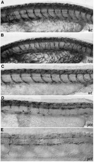
Effect of fss and yot mutations on the primary motoneurons. (A,B) Znp-1 staining of wild-type sibling and fss mutant, respectively. The number of CaP axons is slightly increased in fss as compared to wild type, and some axons terminate prematurely. (C,D) Znp-1 staining of wild-type sibling and yot mutant, respectively. In C, CaP axons project down in every segment. In D only four CaP axons are projecting down. (E) Same yot mutant as in D; high power magnification of five myotomes anterior to the anus. One CaP axon projects into the muscle. In the other segments axons are running posteriorly, parallel to the neural tube. |
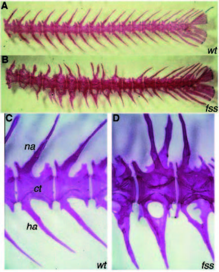
Skeletal phenotype of fss mutants. Skeletal staining on adult wild type (A) and fss mutant (B) tail vertebrae. (C,D) Enlargement of wild-type and mutant vertebrae, respectively at a similar A/P level. ct; centrum, na; neural arch, ha; hemal arch. The centra in fss are almost normal in number and shape, but the length of individual centra is slightly more variable (B). In the wild type (C) every vertebra has four half arches, forming the neural arch on the dorsal side and the hemal arch on the ventral side. In fss mutants (B,D), arches grow out irregularly. For example, on the ventral side of the middle vertebra in D, three half arches are present, two at the left side which fuse, project to the right side, and fuse with the third half arch. PHENOTYPE:
|
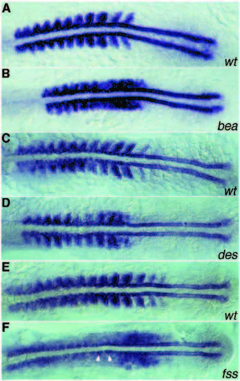
myoD expression in bea, des and fss mutants. All mutants show normal adaxial expression. Wild-type sibling (A) and bea mutant (B), 12-somite stage. In the wild type, striped expression of myoD is visible in the posterior part of every somite. In bea, striped expression is visible in the anteriormost but not in more posterior paraxial mesoderm. (C,D) Wild-type sibling and des mutant, respectively, 13-somite stage. In des, expression in the posterior cells of the first seven somites is normal (D). More posteriorly in the paraxial mesoderm, the stripes change into irregular patches. (E,F) Wild-type sibling and fss mutant, respectively. In fss the striped pattern of expression is no longer seen. Note the elongated adaxial cells just anterior to the region where myoD reaches the highest level of expression (arrowheads). EXPRESSION / LABELING:
PHENOTYPE:
|
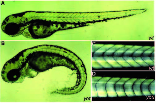
Overview of the phenotype of a wild-type sibling (A) and a yot mutant (B) at 72 hpf. The horizontal myoseptum is visible in A as a black line running through the middle of the myotome. In B this line is absent. yot mutants have a variably curved tail. The swelling of the heart cavity is probably a consequence of the reduced circulation in the tail. Branchial arches are displaced ventrally in yot. This is observed in all mutants where the eyes lie closer together. (C,D) Dark-field image of wild-type sibling (C) and a you mutant (D) at 120 hpf. Myotomes in you are U-shaped instead of V-shaped, and lack the horizontal myoseptum. PHENOTYPE:
|
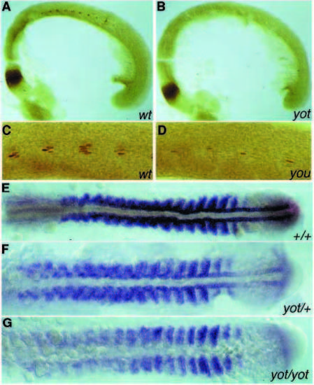
(A) Engrailed (4D9) staining of a 21-somite stage wild-type embryo. Staining is visible at the midbrain-hindbrain boundary and in the somites. In every somite about 2-5 nuclei, representing the muscle pioneers, are stained. In a yot mutant at the same stage (B), staining of the muscle pioneers is absent. (C) 4D9 staining of a wildtype sibling and (D) the weakest you-type mutant, youtm146c. Approximately one Engrailed-positive cell is present per segment. (E-G) myoD-staining of a wild type, a yot mutant and a putative yot/+ embryo at the 17-somite stage, respectively. In the homozygous yot mutant, adaxial staining is absent and the staining in the rest of the paraxial mesoderm is reduced (G). In the yot heterozygous embryo, expression in the adaxial cells is reduced, especially in the presomitic mesoderm, anterior to the tailbud (F). EXPRESSION / LABELING:
PHENOTYPE:
|
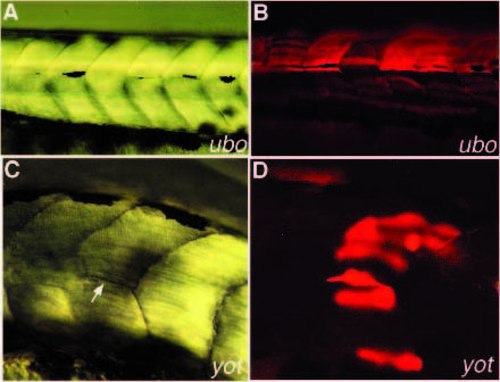
Rescue of the horizontal myoseptum phenotype in ubo and yot mutants by cell transplantation. Rhodamine dextran-labeled wildtype cells were transplanted into ubo or yot mutants at mid-late blastula stage. Embryos were scored for rescue of the myoseptum at 48-72 hpf. (A) Normarski picture of five segments with a rescued myoseptum in an ubo mutant. Note that the segment in the middle does not show any rescue. (B) Picture of the same embryo showing the fluorescently labeled wild type cells. In all segments with rescue, labeled cells are present in the myoseptum. In the myotome that did not show rescue only muscle fibers, that are slightly above or far below the myoseptum, are labeled. (C) Normarski picture of a yot mutant with a single rescued horizontal myoseptum (arrow). (D) Picture of the same region showing a brightly labeled wild-type cell at the position where the myoseptum formed. |

ZFIN is incorporating published figure images and captions as part of an ongoing project. Figures from some publications have not yet been curated, or are not available for display because of copyright restrictions. PHENOTYPE:
|