
FIGURE
Fig. 4
Fig. 4
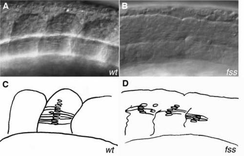
Adaxial cells differentiate normally in fss mutants. (A) Somite 7 at the 13-somite stage, lateral view. At the middle of the somite a stack of flattened nuclei are visible, representing adaxial cells that have elongated and intercalated. (B) fss mutant at a similar region. Nuclear stacks can be seen, although they usually contain less cells. At the point where these cells seem to end a border is forming. (C,D) Schematic drawings of A and B, respectively. |
Expression Data
Expression Detail
Antibody Labeling
Phenotype Data
| Fish: | |
|---|---|
| Observed In: | |
| Stage: | 10-13 somites |
Phenotype Detail
Acknowledgments
This image is the copyrighted work of the attributed author or publisher, and
ZFIN has permission only to display this image to its users.
Additional permissions should be obtained from the applicable author or publisher of the image.
Full text @ Development