|
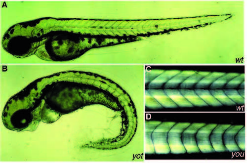
Overview of the phenotype of a wild-type sibling (A) and a yot mutant (B) at 72 hpf. The horizontal myoseptum is visible in A as a black line running through the middle of the myotome. In B this line is absent. yot mutants have a variably curved tail. The swelling of the heart cavity is probably a consequence of the reduced circulation in the tail. Branchial arches are displaced ventrally in yot. This is observed in all mutants where the eyes lie closer together. (C,D) Dark-field image of wild-type sibling (C) and a you mutant (D) at 120 hpf. Myotomes in you are U-shaped instead of V-shaped, and lack the horizontal myoseptum.
|