
|
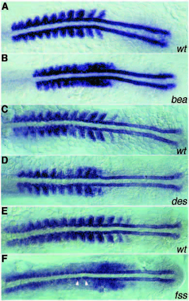
Fig. 7 myoD expression in bea, des and fss mutants. All mutants show normal adaxial expression. Wild-type sibling (A) and bea mutant (B), 12-somite stage. In the wild type, striped expression of myoD is visible in the posterior part of every somite. In bea, striped expression is visible in the anteriormost but not in more posterior paraxial mesoderm. (C,D) Wild-type sibling and des mutant, respectively, 13-somite stage. In des, expression in the posterior cells of the first seven somites is normal (D). More posteriorly in the paraxial mesoderm, the stripes change into irregular patches. (E,F) Wild-type sibling and fss mutant, respectively. In fss the striped pattern of expression is no longer seen. Note the elongated adaxial cells just anterior to the region where myoD reaches the highest level of expression (arrowheads).