
- Title
-
Cell-autonomous shift from axial to paraxial mesodermal development in zebrafish floating head mutants
- Authors
- Halpern, M.E., Thisse, C., Ho, R.K., Thisse, B., Riggleman, B., Trevarrow, B., Weinberg, E.S., Postlethwait, J.H., and Kimmel, C.B.
- Source
- Full text @ Development
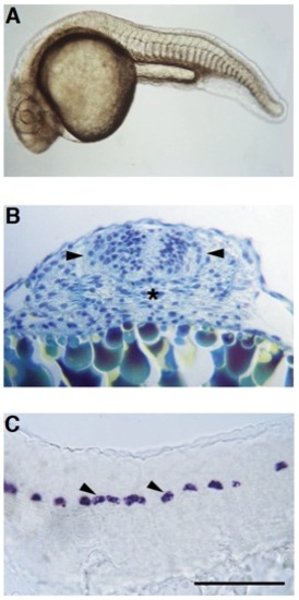
flh mutants have fused somites and discontinuous floor plate. (A) Live flh- pharyngula (approximately 24 hours) with fused somites in the midline. (B) Transverse section through the trunk region of a 24 hour mutant stained with methylene blue-azure II and basic fuchsin (Humphrey and Pittman, 1974) shows differentiating muscle fibers across the midline. The developing spinal cord (arrowheads), and the position where the notochord would normally be located (asterisk), are indicated. (C) Scattered patches of floor plate cells (arrowheads) in the trunk and tail spinal cord of a 24 hour flh mutant are revealed by in situ hybridization with a probe for a-collagen II, which normally is expressed in the floor plate, notochord and hypochord (Yan et al., 1995). Scale bar, 100 μm for B and C. |
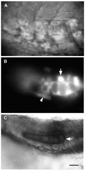
WT-derived notochord can differentiate in flh mutant hosts. (A) Side view of a live 24 hour flh mutant that differentiated a short stretch (8 cells) of morphologically identifiable notochord following cell transplantation at the blastula stage. (A) Nomarski optics and (B) the corresponding fluorescent image. (B) Notochord cells developed from rhodamine-dextran labeled WT donor cells. Labeled WT cells also gave rise to floor plate above the notochord (arrow) and hypochord cells (arrowhead) beneath it. (C) A subset of the transplanted WT cells (arrow) stained with MZ15 (Smith and Watt, 1985), an anti-keratan sulfate monoclonal antibody specific for notochord. MZ15 also labels floor plate and the spinal cord central canal (Hatta, 1992). Scale bar, 20 μm for A and B; 30 μm for C. |
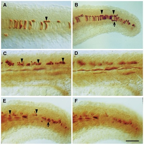
Dorsal axial transplants of flh mutant cells differentiate into floor plate and muscle instead of floor plate and notochord. Sagittal views of 24 hour WT host embryos with (A, B) WT or (C-F) flh mutant donor cells (dark brown) derived from dorsal axial transplants. A transplant was defined as a dorsal axial transplant if donor cells contributed to >20 floor plate cells in the ventral spinal cord of the trunk and tail. (A,B) In the case of control experiments with WT cells, donor cells that gave rise to floor plate (arrowheads), also gave rise to notochord cells (93% of dorsal axial transplants, n=27/29, present study and 95%, n=56/59, Halpern et al., 1993), and sometimes hypochord cells (arrow in B). (C) flh- donor cells contributed to floor plate (arrowheads) but not notochord. (D) Instead, mutant cells formed muscle fibers. A total of 72 blastulas were transplanted. Out of 17 WT embryos receiving flh- cells, 7 had dorsal axial transplants. Of these, mutant donor cells gave rise to more than 50 muscle fibers in 5 embryos and approximately 35 fibers in 1 embryo. Mutant muscle fibers were often found in the middle of the myotome, with respect to the dorsoventral axis, at the position of muscle pioneers (Felsenfeld et al., 1991). Although flh- cells did not contribute to WT hypochord, due to the lower frequency of donor cells forming hypochord in control transplants, more flh genetic mosaics are needed to confirm that flh- cells are unable to form hypochord. (E,F) Mutant donor cells produced muscle fibers in newly formed somites, however, undifferentiated mesodermal cells in more posterior, and hence, developmentally younger, regions were situated in the midline at the level of the WT notochord (arrow). (C,D, and E,F) Different focal planes of the same embryos. Scale bar = 50 μm for A, C, D, E and F; 75 μm for B. PHENOTYPE:
|
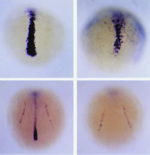
Axial mesodermal differentiation is initiated but not maintained in flh mutants. twist is expressed by axial mesoderm in (A) WT and (B) flh mutant gastrulas at 80% epiboly (8.25 hours). At the 3-somite stage (11 hours) in (C) WT and (D) mutant embryos, twist is also expressed in the presumptive pronephric ducts. Although twist transcripts are abundant in newly-forming WT notochord at this stage (C), expression is entirely absent in the flh- axis (D). In B, flh mutant embryos were overstained approximately three-fold relative to WT sibling embryos. Overstaining of flh- embryos at early somite stages comparable to D, failed to reveal twist transcripts in the axis. EXPRESSION / LABELING:
|
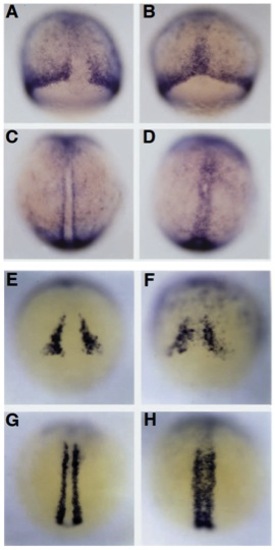
flh- axial cells express markers of paraxial mesoderm. At 75% epiboly (8 hours) in (A) WT embryos, snail1 is expressed in the marginal region and in mesodermal cells flanking the dorsal axis, and in (B) flh mutants, snail1 is also ectopically expressed by cells in the axis. At the 3-somite stage (11 hours) in (C) WT embryos, expression persists in paraxial mesoderm and also in (D) axial cells in flh mutants. Zebrafish myoD is only expressed in paraxial mesodermal cells flanking the dorsal axis and not throughout the margin in 60% epiboly (E) WT embryos. Dorsal expression is mostly absent, however, a small number of cells in the (F) flh mutant axis (60% epiboly) contain myoD transcripts. By early somitogenesis (3-5 somites), when myoD is strongly expressed in (G) WT paraxial mesoderm, many more cells in (H) the flh mutant axis also express myoD. In other strongly stained preparations, myo D expression was also found more laterally in the developing somites (data not shown). EXPRESSION / LABELING:
|
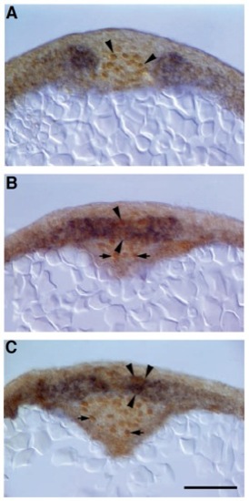
Single cells in the flh mutant midline co-express axial and paraxial mesodermal genes. Transverse sections (7 mm) of 2-somite stage (10J hours) (A) WT and (B,C) flh mutant embryos double labeled for Ntl protein and snail1 RNA expression. (A) In the WT axis, nuclei of presumptive notochord cells express Ntl (arrowheads), while snail1 expression is confined to the paraxial presomitic mesoderm. (B,C) Single cells (arrowheads) in the flh mutant axis express Ntl protein (brown nucleus) and snail1 transcripts (blue cytoplasm). (B) A more rostral section through the same embryo as C. Other cells expressing Ntl protein in B and C (arrows), constitute an epithelium surrounding Kupffer’s vesicle (see Laale, 1985), an axial structure at the tip of the newly forming notochord in WT embryos (data not shown). Although axial mesodermal cells express paraxial mesodermal genes in flh mutants, cells lining Kupffer’s vesicle only express Ntl protein as normal. Scale bar, 50 μm. EXPRESSION / LABELING:
PHENOTYPE:
|