
|
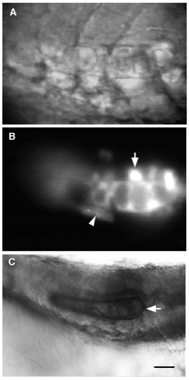
Fig. 2 WT-derived notochord can differentiate in flh mutant hosts. (A) Side view of a live 24 hour flh mutant that differentiated a short stretch (8 cells) of morphologically identifiable notochord following cell transplantation at the blastula stage. (A) Nomarski optics and (B) the corresponding fluorescent image. (B) Notochord cells developed from rhodamine-dextran labeled WT donor cells. Labeled WT cells also gave rise to floor plate above the notochord (arrow) and hypochord cells (arrowhead) beneath it. (C) A subset of the transplanted WT cells (arrow) stained with MZ15 (Smith and Watt, 1985), an anti-keratan sulfate monoclonal antibody specific for notochord. MZ15 also labels floor plate and the spinal cord central canal (Hatta, 1992). Scale bar, 20 μm for A and B; 30 μm for C.