
|
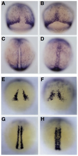
Fig. 5 flh- axial cells express markers of paraxial mesoderm. At 75% epiboly (8 hours) in (A) WT embryos, snail1 is expressed in the marginal region and in mesodermal cells flanking the dorsal axis, and in (B) flh mutants, snail1 is also ectopically expressed by cells in the axis. At the 3-somite stage (11 hours) in (C) WT embryos, expression persists in paraxial mesoderm and also in (D) axial cells in flh mutants. Zebrafish myoD is only expressed in paraxial mesodermal cells flanking the dorsal axis and not throughout the margin in 60% epiboly (E) WT embryos. Dorsal expression is mostly absent, however, a small number of cells in the (F) flh mutant axis (60% epiboly) contain myoD transcripts. By early somitogenesis (3-5 somites), when myoD is strongly expressed in (G) WT paraxial mesoderm, many more cells in (H) the flh mutant axis also express myoD. In other strongly stained preparations, myo D expression was also found more laterally in the developing somites (data not shown).