
|
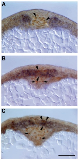
Fig. 6 Single cells in the flh mutant midline co-express axial and paraxial mesodermal genes. Transverse sections (7 mm) of 2-somite stage (10J hours) (A) WT and (B,C) flh mutant embryos double labeled for Ntl protein and snail1 RNA expression. (A) In the WT axis, nuclei of presumptive notochord cells express Ntl (arrowheads), while snail1 expression is confined to the paraxial presomitic mesoderm. (B,C) Single cells (arrowheads) in the flh mutant axis express Ntl protein (brown nucleus) and snail1 transcripts (blue cytoplasm). (B) A more rostral section through the same embryo as C. Other cells expressing Ntl protein in B and C (arrows), constitute an epithelium surrounding Kupffer’s vesicle (see Laale, 1985), an axial structure at the tip of the newly forming notochord in WT embryos (data not shown). Although axial mesodermal cells express paraxial mesodermal genes in flh mutants, cells lining Kupffer’s vesicle only express Ntl protein as normal. Scale bar, 50 μm.