|
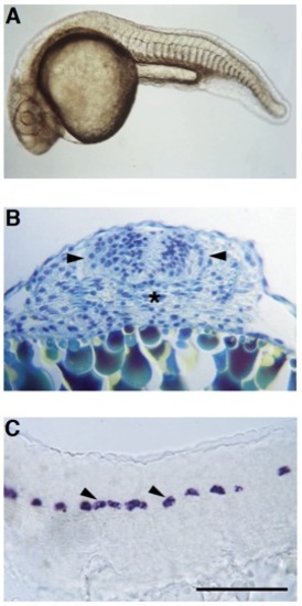
flh mutants have fused somites and discontinuous floor plate. (A) Live flh- pharyngula (approximately 24 hours) with fused somites in the midline. (B) Transverse section through the trunk region of a 24 hour mutant stained with methylene blue-azure II and basic fuchsin (Humphrey and Pittman, 1974) shows differentiating muscle fibers across the midline. The developing spinal cord (arrowheads), and the position where the notochord would normally be located (asterisk), are indicated. (C) Scattered patches of floor plate cells (arrowheads) in the trunk and tail spinal cord of a 24 hour flh mutant are revealed by in situ hybridization with a probe for a-collagen II, which normally is expressed in the floor plate, notochord and hypochord (Yan et al., 1995). Scale bar, 100 μm for B and C.
|