- Title
-
Second-order projection from the posterior lateral line in the early zebrafish brain
- Authors
- Fame, R.M., Brajon, C., and Ghysen, A.
- Source
- Full text @ Neural Dev.
|
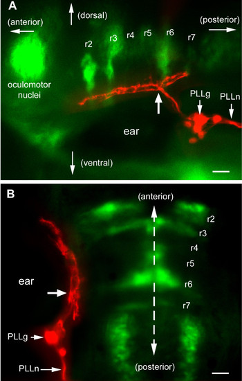
Central projection of PLL sensory neurons in an islet-GFP background, as seen (a) laterally and (b) dorsally. Rhombomere positions are based on the morphology of the various motor nuclei, particularly trigeminal in r2 and r3, facial in r6, and in r7 the small lateral line caudal efferent nucleus [33]. The exit of the facial nerve in r4, barely visible in (a), provides an additional landmark. DiI has been injected in the PLL nerve (PLLn). The cell bodies of the sensory neurons are gathered in the PLL ganglion (PLLg). Their axons enter the hindbrain posterior to the otic vesicle (ear) and bifurcate at the level of r6 (site of bifurcation marked by a thick arrow in both panels). They extend anteriorly to the level of r1 and posteriorly to the level of r7. The red and green images were taken at different focal planes. In this and in all following figures the dashed line indicates the sagittal plane and the scale bar is 20 microns. |
|
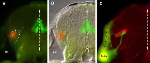
Transversal sections of the afferent projection. At the level of r3–r4, the PLL projection extends at the lateral edge of the hindbrain, while the basal-plate derived motor neurons are present near the midline. Based on (a) autofluorescence and (b) the Nomarski image, the afferent projection extends within a neuropilic region (dotted line). (c) At a slightly more anterior level, the PLL projection labeled with DiI (red) and the ALL projection labeled with DiO (green) are apposed yet segregated. Due to the thickness of the vibratome section (100 micorns), the same section also comprises the ALL ganglion (ALLg) and shows contaminating labeling of the hindbrain surface (asterisks) close to the site of DiO injection. |
|
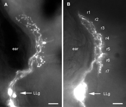
Dorsal aspect of the first-order PLL projection. Dye injection was done either in (a) a single neuromast, resulting in the specific labeling of the two neurons that innervate this neuromast or (b) in the PLL nerve, resulting in the labeling of many PLL sensory neurons. In both cases the axons outline a well-defined and compact synaptic field. The projection in (b) was observed in an islet-GFP background, allowing us to identify rhombomeres. |
|
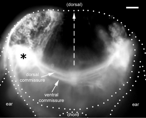
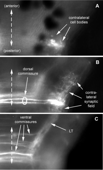
Distribution of second-order neurons. A vibratome section at the level of the site of DiI injection (asterisk) reveals a large number of ipsilateral cell bodies, a more limited number of contralateral cell bodies, and two commissures connecting the left and right lateral line nuclei. The impression of massive spread of DiI is due to the saturation effect of scattered fluorescent light; a lower exposure reveals that the injection was confined to a much smaller region than the white blob in the figure. In this and all subsequent figures, the left PLL synaptic field has been labeled with DiI (in those cases where the injection was on the right, the figures have been inverted to simplify the perception by the reader). |
|
Dorsal aspect of the commissures as seen at three focal levels. From dorsal to ventral: contralateral neurons labeled after injection of DiI in the left PLL synaptic field (a) have their cell bodies located at a dorsal level, and (b) send their axons along a dorsal commissure and their dendrites invade the contralateral synaptic field. Ipsilateral neurons extending their axon to the contralateral hindbrain nucleus presumably use the dorsal commissure as well (b). (c) Other ipsilateral neurons send their axons through more ventral commissures and form the LT branch. All images (a-c) are composites of two consecutive focal planes within an extended Z-series. |
|
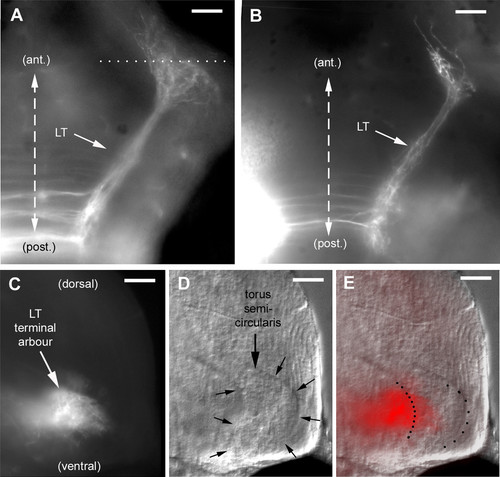
The LT branch extends to the torus semicircularis. (a, b) As seen dorsally in whole-mount embryos, the LT projection courses anteriorly and laterally at a level slightly ventral to the ventral commissures, and turns abruptly upon reaching its target (see also Figures 7 and 12). In (a), the dotted line indicates the approximative plane of the vibratome section shown in (c-e). (c) In this section, the terminal arbor of the LT projection is seen to extend along the torus semicircularis, (d) which is easily delineated under Nomarski optics. (c, e) Fine fibers from the LT arbor extend into the torus. |
|
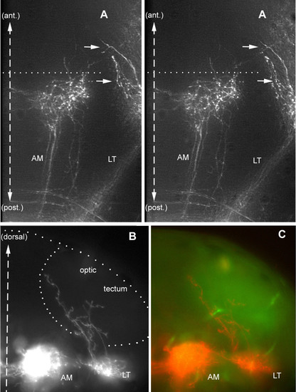
A few fibers escape from the LT projection and climb into the optic tectum. (a) Dorsal stereo-view of a whole-mount embryo (to be seen with crossed eyes). The lettering has been adjusted to correspond to the dorso-ventral level of the corresponding features; thus, the arrows that point to the climbing fibers will appear much more dorsal to 'AM', whereas 'LT' will appear slightly ventral to 'AM'. (b, c) Climbing fibers seen in a vibratome section. The outline of the tectum in (b) has been drawn based on the autofluorescence pattern seen under blue exciting light (c), which reveals fibrous material (neuropil) as opposed to cell bodies (nuclei). |
|
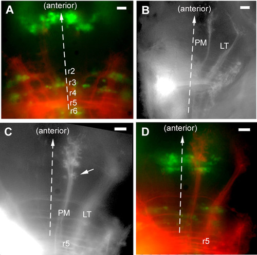
Other posterior projections. (a) A projection that was observed in three out of five cases when the site of injection was deliberately out of the PLL synaptic field, slightly medial to it. (b) Overall view of the projection resulting from injections in the posterior region of the synaptic field, illustrating the LT projection and posterior medial (PM) projection. (c, d) The PM projection extends through the ocular motor complex (oculomotor and trochlear nuclei) and arborizes both posterior (arrow in (c)) and anterior to this cluster. |
|
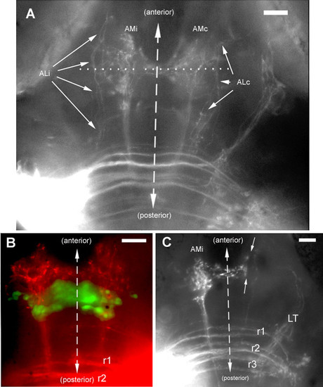
Projections from the anterior PLL synaptic field. (a) The ipsi- and contralateral anterior medial projections (AMi, AMc) course through well-defined symmetrical tracts, the MLF (median longitudinal fascicle), and extend very dense commissural fibers. In contrast, the anterior lateral fibers (ALi, ALc) are but loosely fasciculated (arrows). (b) The AM projection extends through the ocular motor nuclei and overlaps partly with the PM projection but it differs from the latter in being bilateral with commissural connections, and in extending further anteriorly and laterally. (c) The commissural fibers do not invade the contralateral projection but stop sharply at its boundary (arrows) |
|
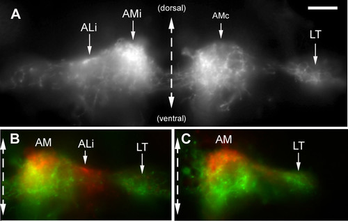
Lateralization of the AM branches. (a) Transversal section through the arborization of the AM branches reveals that the ipsilateral projection is very dense in the mediodorsal region of the target area, while the contralateral projection extends more ventrally within the same area. (b, c) This difference was emphasized by superimposing the ipsilateral half of the section (colored in red) over the contralateral one (colored in green) for sections of two different embryos. As expected, the LT branch is mostly green, consistent with its contralateral predominance. In (b), the ALi branch happens to be more labeled than ALc and appears, therefore, in red. |
|
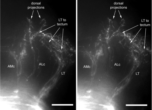
Dorsal extensions of the ALc branch. Stereo-view of a whole mount embryo illustrating the dorsal fibers emanating from the ALc. Lettering emphasizes the dorso-ventral level of the corresponding features, as in Figure 7. When seen with crossed eyes, this view will reveal two fibers climbing from the tip of the ALc projection, slightly anterior to the fibers that extend from the LT projection into the tectum (labeled 'LT to tectum'). The ALc fibers do not reach a level as dorsal as the LT fiber ramification, consistent with a target in the pretectal region of the embryonic brain. |
|
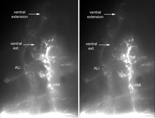
Ventral extension of the ALi branch. Stereo-view illustrating the fibers that extend ventrally from the ALi. One fiber extends ventral to the AMi projection, and more ventral extensions are also observed at more anterior levels. |
|
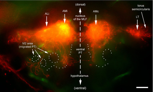
Ventral extension of ALi. Transversal section cut at the level of the commissural fibers (dotted line in Figure 10a). Two prominent ventral extensions are observed. A medial extension is adjacent to (and crosses) the posterior tuberculum (PT), a thalamic derivative. A more lateral extension arborizes in the M2 area. This region will form the preglomerular nuclei, the major diencephalic target of third-order lateral line projection in the adult. The outline of the major nuclei (dotted line) is based on autofluorescence, which reveals fibrous material (neuropils) as opposed to cell bodies (nuclei). |
|
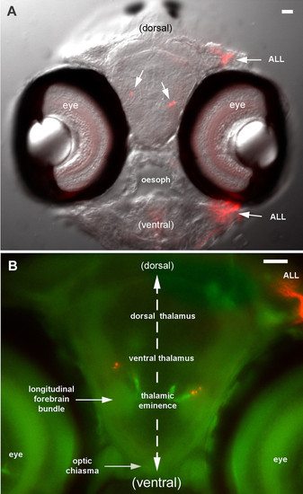
Anterior extensions of ALc and ALi. (a) Transverse section of the head at the level of the optic chiasma. The injection in the PLL synaptic field extended to the ALL synaptic field such that ALL neuromasts and nerves are also labeled (ALL). (b) Position of the labeled fibers in relation to the major forebrain subdivisions, based on the autofluorescence pattern (green). Since the thick section is subjectively seen from the front, the injection site appears on the right side of this figure (left side of the fish). |
|
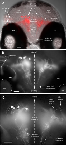
Anterior neurons projecting back to the hindbrain. (a) Transverse section revealing the overall pattern at the level of the postotic commissure. A large amount of DiI was injected, resulting in the back-labeling of ALL neurons on the ipsilateral side as well as of central neurons at the midbrain-forebrain boundary. (b) Higher magnification revealing fibers that follow the post-optic commissure. Since such fibers are only observed when back-projecting neurons are labeled, we believe that they belong to the latter. (c) Detail of the same preparation cleaned with the 'Rapid Deconvolution' program of IPLab (see Materials and methods) to enhance the cell bodies of the back-projecting neurons. Also present on the contralateral side of this section is a fiber of ALc extending dorsally, possibly into the pretectum, as well as the anteriormost branchlet of a LT fiber that extends into the optic tectum. |

