
FIGURE
Fig. 14
- ID
- ZDB-FIG-061228-22
- Publication
- Fame et al., 2006 - Second-order projection from the posterior lateral line in the early zebrafish brain
- Other Figures
- All Figure Page
- Back to All Figure Page
Fig. 14
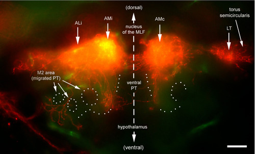
Ventral extension of ALi. Transversal section cut at the level of the commissural fibers (dotted line in Figure 10a). Two prominent ventral extensions are observed. A medial extension is adjacent to (and crosses) the posterior tuberculum (PT), a thalamic derivative. A more lateral extension arborizes in the M2 area. This region will form the preglomerular nuclei, the major diencephalic target of third-order lateral line projection in the adult. The outline of the major nuclei (dotted line) is based on autofluorescence, which reveals fibrous material (neuropils) as opposed to cell bodies (nuclei). |
Expression Data
Expression Detail
Antibody Labeling
Phenotype Data
Phenotype Detail
Acknowledgments
This image is the copyrighted work of the attributed author or publisher, and
ZFIN has permission only to display this image to its users.
Additional permissions should be obtained from the applicable author or publisher of the image.
Full text @ Neural Dev.