
Fig. 4
- ID
- ZDB-FIG-061228-12
- Publication
- Fame et al., 2006 - Second-order projection from the posterior lateral line in the early zebrafish brain
- Other Figures
- All Figure Page
- Back to All Figure Page
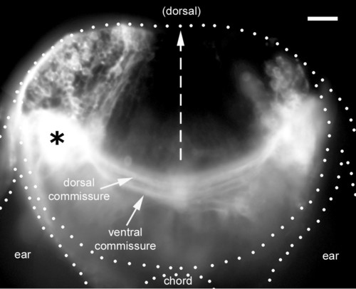
Distribution of second-order neurons. A vibratome section at the level of the site of DiI injection (asterisk) reveals a large number of ipsilateral cell bodies, a more limited number of contralateral cell bodies, and two commissures connecting the left and right lateral line nuclei. The impression of massive spread of DiI is due to the saturation effect of scattered fluorescent light; a lower exposure reveals that the injection was confined to a much smaller region than the white blob in the figure. In this and all subsequent figures, the left PLL synaptic field has been labeled with DiI (in those cases where the injection was on the right, the figures have been inverted to simplify the perception by the reader). |