
Fig. 5
- ID
- ZDB-FIG-061228-14
- Publication
- Fame et al., 2006 - Second-order projection from the posterior lateral line in the early zebrafish brain
- Other Figures
- All Figure Page
- Back to All Figure Page
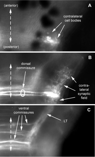
Dorsal aspect of the commissures as seen at three focal levels. From dorsal to ventral: contralateral neurons labeled after injection of DiI in the left PLL synaptic field (a) have their cell bodies located at a dorsal level, and (b) send their axons along a dorsal commissure and their dendrites invade the contralateral synaptic field. Ipsilateral neurons extending their axon to the contralateral hindbrain nucleus presumably use the dorsal commissure as well (b). (c) Other ipsilateral neurons send their axons through more ventral commissures and form the LT branch. All images (a-c) are composites of two consecutive focal planes within an extended Z-series. |