
|
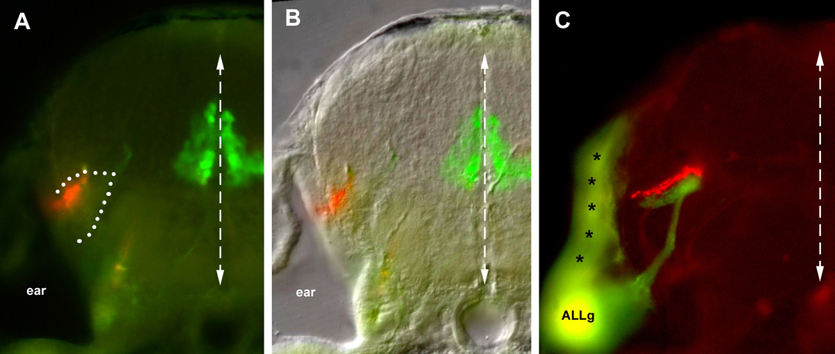
Fig. 2 Transversal sections of the afferent projection. At the level of r3–r4, the PLL projection extends at the lateral edge of the hindbrain, while the basal-plate derived motor neurons are present near the midline. Based on (a) autofluorescence and (b) the Nomarski image, the afferent projection extends within a neuropilic region (dotted line). (c) At a slightly more anterior level, the PLL projection labeled with DiI (red) and the ALL projection labeled with DiO (green) are apposed yet segregated. Due to the thickness of the vibratome section (100 micorns), the same section also comprises the ALL ganglion (ALLg) and shows contaminating labeling of the hindbrain surface (asterisks) close to the site of DiO injection.