
|
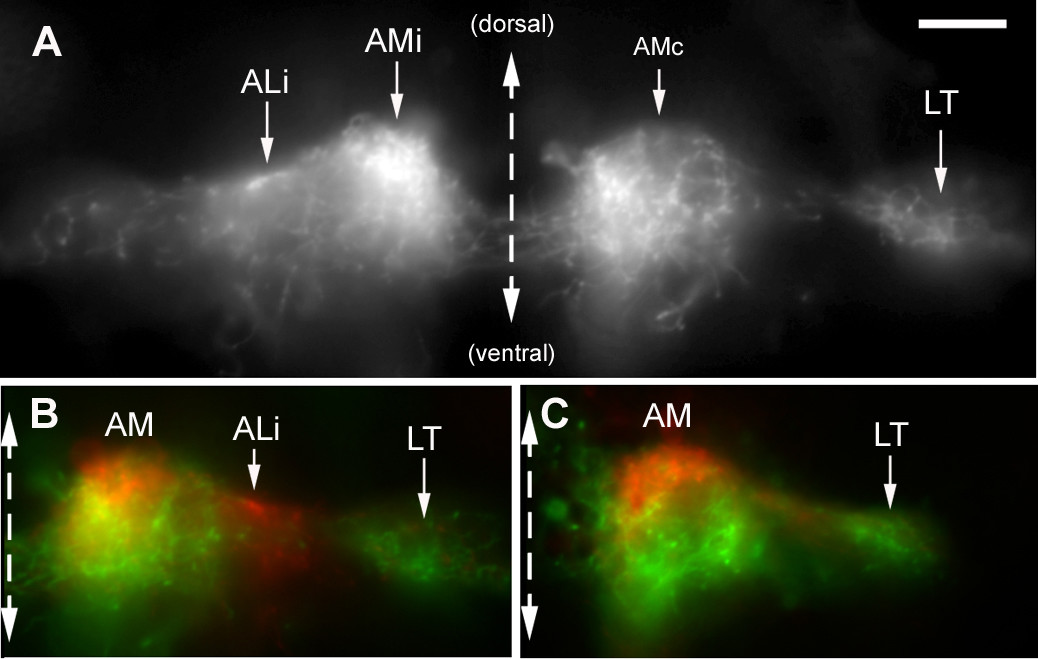
Fig. 11 Lateralization of the AM branches. (a) Transversal section through the arborization of the AM branches reveals that the ipsilateral projection is very dense in the mediodorsal region of the target area, while the contralateral projection extends more ventrally within the same area. (b, c) This difference was emphasized by superimposing the ipsilateral half of the section (colored in red) over the contralateral one (colored in green) for sections of two different embryos. As expected, the LT branch is mostly green, consistent with its contralateral predominance. In (b), the ALi branch happens to be more labeled than ALc and appears, therefore, in red.