
Image
|
Figure Caption
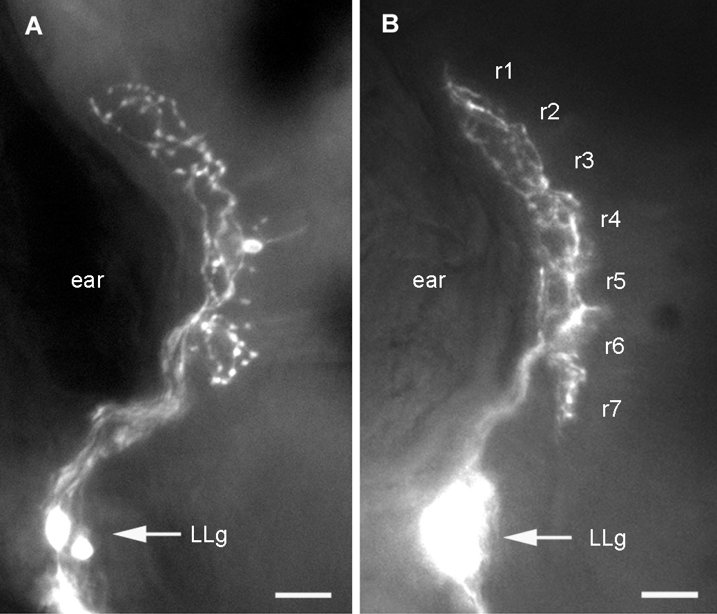
Fig. 3 Dorsal aspect of the first-order PLL projection. Dye injection was done either in (a) a single neuromast, resulting in the specific labeling of the two neurons that innervate this neuromast or (b) in the PLL nerve, resulting in the labeling of many PLL sensory neurons. In both cases the axons outline a well-defined and compact synaptic field. The projection in (b) was observed in an islet-GFP background, allowing us to identify rhombomeres.
Acknowledgments
This image is the copyrighted work of the attributed author or publisher, and
ZFIN has permission only to display this image to its users.
Additional permissions should be obtained from the applicable author or publisher of the image.
Full text @ Neural Dev.