
- Title
-
Semaphorin 3d promotes cell proliferation and neural crest cell development downstream of TCF in the zebrafish hindbrain
- Authors
- Berndt, J.D., and Halloran, M.C.
- Source
- Full text @ Development
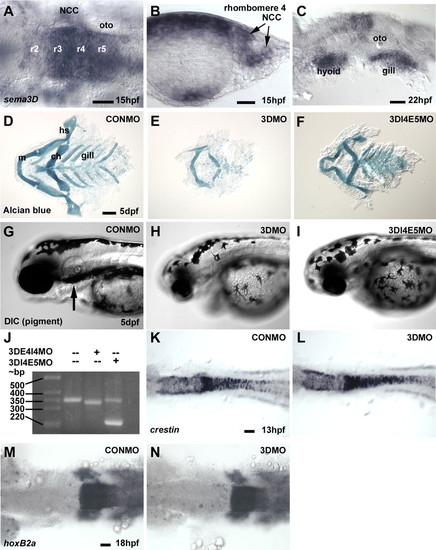
Sema3d mRNA is expressed in NCCs and morpholino knockdown of Sema3d disrupts NCC derivatives without affecting NCC induction. (A-C) In situ hybridization for sema3d at 15 hpf (A,B) and 22 hpf (C). (D-F) Alcian Blue labeling of pharyngeal cartilage at 5 dpf; (G-I) DIC imaging of melanophores in 2 dpf in CONMO (D,G), 3DMO (E,H) and 3DI4E5MO (F,I) injected embryos. Arrow in G indicates ventral horn of melanophores. (J) RT-PCR on mRNA extracted from embryos injected with Sema3d splice blocking morpholinos. (K-N) In situ hybridization for crestin at 13 hpf (K,L) and hoxb2a at 18 hpf (M,N) in CONMO (K,M) and 3DMO (L,N) injected embryos. (A,K-N) Dorsal views, anterior is leftwards. (B) Transverse section through rhombomere 4. (C,G-I) Lateral views, anterior is leftwards. (D-F) Ventral views, anterior is leftwards. r2-r5, rhombomeres 2-5; oto, otocyst; m, hs, ch and gill, Meckel's, hyosymplectic, ceratohyal and gill cartilages, respectively; bp, base pairs. Scale bars: 40 μm for A,C,M,N; 20 μm for B; 100 μm for D-I; 80 μm for K,L. EXPRESSION / LABELING:
|
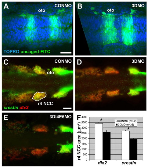
Sema3d knockdown reduces migratory NCC markers without affecting EMT. (A,B) Uncaged FITC-dextran (green) and TOPRO3 labeling of nuclei (blue) in the hindbrain at 17 hpf in CONMO (A) and 3DMO (B) injected embryos. (C-E) Double in situ hybridization for crestin (green) and dlx2 (red); expression is reduced in 3DMO (D) and 3DI4E5MO (E) injected embryos compared with CONMO (C). (F) Quantification of the area of labeling of crestin and dlx2 in the r4 migratory stream, indicated by white outline in C. *P<10-6, t-test. (A-E) Dorsal views, anterior is leftwards. oto, otocyst. Scale bars: 40 μm. EXPRESSION / LABELING:
|
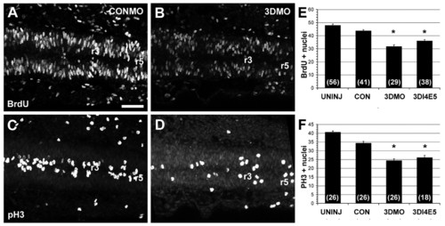
Sema3d knockdown inhibits cell cycle in the hindbrain. (A-D) Immunodetection in the hindbrain at 17 hpf of BrdU (A,B) and PH3 (C,D) in CONMO (A,C) and 3DMO (B,D) injected embryos. (E,F) Quantification of the number of BrdU (E) and PH3 (F) labeled nuclei in the neuroepithelium of rhombomeres 3-5. Numbers in parentheses indicate n. *P<10-4 compared with CONMO, t-test. (A-D) Dorsal views, anterior is leftwards. Scale bar: 40 μm for A-D. |
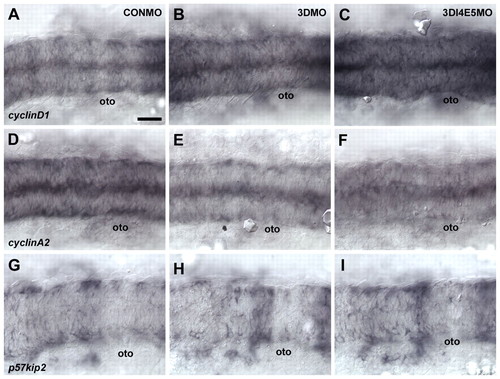
Sema3d knockdown affects the expression of cell cycle genes. (A-I) In situ hybridization in the hindbrain at 17hpf for cyclin D1 (A-C), cyclin A2 (D-F) and p57kip2 (G-I) in CONMO (A,D,G), 3DMO (B,E,H), and 3DI4E5MO (C,F,I) injected embryos. oto, otocyst. (A-I) Dorsal views, anterior is leftwards. Scale bars: 40 μm for A-I. EXPRESSION / LABELING:
|
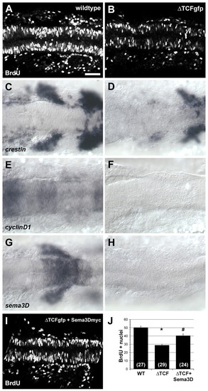
ΔTCFgfp disrupts cell cycle and crestin expression and eliminates cyclin D1 and sema3d expression. (A-I) Immunodetection of BrdU (A,B,I) and in situ hybridization for crestin (C,D), cyclinD1 (E,F), sema3d (G,H) in the hindbrain at 17 hpf of heat-shocked wild-type siblings (A,C,E,G), ΔTCFgfp transgenic (B,D,F,H), and ΔTCFgfp + Sema3dmyc double-transgenic (I) embryos. (J) Quantification of the number of BrdU-labeled nuclei in the neuroepithelium of rhombomeres 3-5. Numbers in parentheses indicate n. *P<10-13 versus wild-type sibling, #P<10-4 versus ΔTCFgfp, t-test. (A-I) Dorsal views, anterior is leftwards. oto, otocyst. Scale bar: 40 μm for A-I. EXPRESSION / LABELING:
|
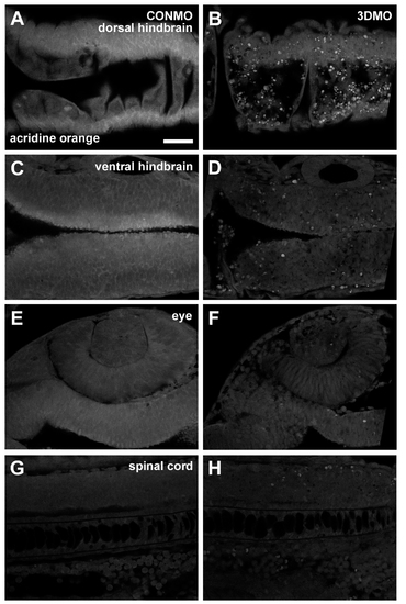
Cell death is increased specifically in the dorsal neural epithelium following Sema3d knockdown. (A-H) Acridine Orange labeling of cell death at 24 hpf in the dorsal hindbrain (A,B), ventral hindbrain (C,D), eye (E,F) and spinal cord (G,H) in CONMO (A,C,E,G) and 3DMO (at 250 μM) (B,D,F,H) injected embryos. (A-F) Dorsal views, anterior is leftwards. (G,H) Lateral view, anterior is leftwards. Scale bar: 40 μm for A-H. |
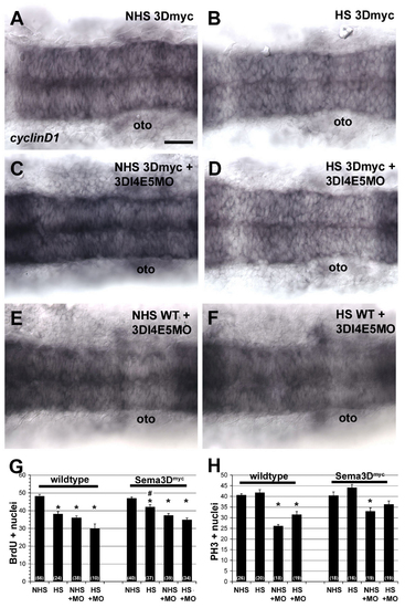
Rescue of the Sema3D splice morpholino (3DI4E5MO) by Sema3Dmyc overexpression. (A-F) In situ hybridization for cyclin D1 in the hindbrain at 17 hpf in Sema3Dmyc transgenic embryos (A-D) or wild-type embryos (E,F) that were uninjected (A,B) or injected with 3DI4E5MO (C-F) and were either not heat-shocked (A,C,E) or heat-shocked for 1 hour at 14 hpf (B,D,F). (G,H) Quantification of the number of BrdU (I) and PH3 (J) labeled nuclei in the neuroepithelium of rhombomeres 3-5. Numbers in parentheses indicate n. *P<0.005 versus uninjected NHS, #P<0.03 versus wild-type HS. oto, otocyst. (A-F) Dorsal views, anterior is leftwards. Scale bars: 40 μm for A-F. EXPRESSION / LABELING:
|

Unillustrated author statements EXPRESSION / LABELING:
|