FIGURE

Fig. S2

ID
ZDB-FIG-061110-7
Publication
Berndt et al., 2006 - Semaphorin 3d promotes cell proliferation and neural crest cell development downstream of TCF in the zebrafish hindbrain
Other Figures
All Figure Page
Back to All Figure Page
Fig. S2

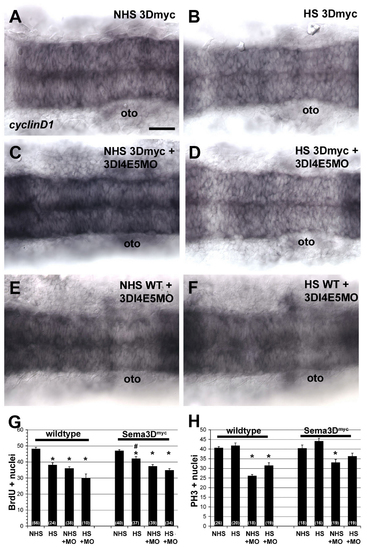
Rescue of the Sema3D splice morpholino (3DI4E5MO) by Sema3Dmyc overexpression. (A-F) In situ hybridization for cyclin D1 in the hindbrain at 17 hpf in Sema3Dmyc transgenic embryos (A-D) or wild-type embryos (E,F) that were uninjected (A,B) or injected with 3DI4E5MO (C-F) and were either not heat-shocked (A,C,E) or heat-shocked for 1 hour at 14 hpf (B,D,F). (G,H) Quantification of the number of BrdU (I) and PH3 (J) labeled nuclei in the neuroepithelium of rhombomeres 3-5. Numbers in parentheses indicate n. *P<0.005 versus uninjected NHS, #P<0.03 versus wild-type HS. oto, otocyst. (A-F) Dorsal views, anterior is leftwards. Scale bars: 40 μm for A-F.

Expression Data
Gene:
Fish:
Condition:
Knockdown Reagent:
Anatomical Terms:
Stage: 14-19 somites

Expression Detail
Antibody Labeling
Phenotype Data

Phenotype Detail
Acknowledgments
This image is the copyrighted work of the attributed author or publisher, and ZFIN has permission only to display this image to its users. Additional permissions should be obtained from the applicable author or publisher of the image. Full text @ Development