
FIGURE
Fig. 2
- ID
- ZDB-FIG-061104-24
- Publication
- Berndt et al., 2006 - Semaphorin 3d promotes cell proliferation and neural crest cell development downstream of TCF in the zebrafish hindbrain
- Other Figures
- All Figure Page
- Back to All Figure Page
Fig. 2
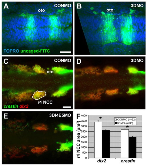
Sema3d knockdown reduces migratory NCC markers without affecting EMT. (A,B) Uncaged FITC-dextran (green) and TOPRO3 labeling of nuclei (blue) in the hindbrain at 17 hpf in CONMO (A) and 3DMO (B) injected embryos. (C-E) Double in situ hybridization for crestin (green) and dlx2 (red); expression is reduced in 3DMO (D) and 3DI4E5MO (E) injected embryos compared with CONMO (C). (F) Quantification of the area of labeling of crestin and dlx2 in the r4 migratory stream, indicated by white outline in C. *P<10-6, t-test. (A-E) Dorsal views, anterior is leftwards. oto, otocyst. Scale bars: 40 μm. |
Expression Data
| Genes: | |
|---|---|
| Fish: | |
| Knockdown Reagents: | |
| Anatomical Term: | |
| Stage: | 14-19 somites |
Expression Detail
Antibody Labeling
Phenotype Data
Phenotype Detail
Acknowledgments
This image is the copyrighted work of the attributed author or publisher, and
ZFIN has permission only to display this image to its users.
Additional permissions should be obtained from the applicable author or publisher of the image.
Full text @ Development