|
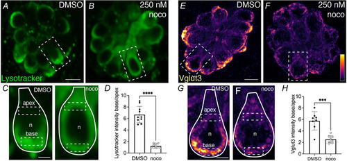
An intact microtubule network is required to enrich LysoTracker and Vglut3 at the presynapse A–C, example live image of LysoTracker Red (green) to label synaptic vesicles in neuromasts at 5 dpf in wild‐type larva treated with 250 nm nocodazole (B) or DMSO control (A). The dashed box in each image indicates the hair cell magnified and outlined with a continuous line in C. D, quantification shows significantly less LysoTracker enrichment at the cell base in nocodazole‐treated larvae compared with DMSO control (control: 6.68 ± 1.43; nocodazole: 1.22 ± 0.37, n = 10 control and nocodazole neuromasts, unpaired t test, P < 0.0001). E–G, example immunolabel of Vglut3 to label synaptic vesicles in neuromasts at 5 dpf in larva treated with 250 nm nocodazole (F) or DMSO control (E). The dashed box in each image indicates the hair cell magnified and outlined with a continuous line in G. H, quantification reveals significantly less Vglut3 enrichment at the cell base in nocodazole‐treated larvae compared with DMSO controls (control: 5.75 ± 1.61; nocodazole: 2.53 ± 0.65, n = 8 control and nocodazole neuromasts, unpaired t test, P < 0.0001). The solid lines in the magnified images outline a single hair cell, with the base of the cell at the bottom of the image, and dashed boxes indicate example ROIs of the apical and basal regions used for quantification. n indicates nucleus. Scale bars in A and E = 5 µm and 2 µm in C and G.
|