|
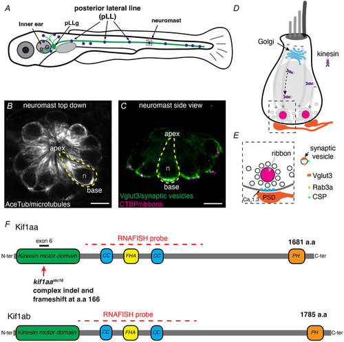
The zebrafish lateral‐line system and Kif1a paralogues A, schematic of a larval zebrafish at 5 dpf. Hair cells are present in the inner ear and lateral line (blue). Hair cells are innervated by neurons in the posterior lateral‐line ganglion (pLLg) located near the inner ear (green). B–C, the lateral line is composed of clusters of hair cells called neuromasts (5 dpf). The apices of hair cells project from the centre of these clusters, while the ribbon synapses are located at the base of the cells. Neuromasts can be viewed from the top down (B) or from the side (C). An individual hair cell in B and C is outlined in yellow. Within hair cells, microtubule networks extend along the apical–basal axis (B). At the base of hair cells, synaptic vesicles (Vglut3 label, green) are enriched near the presynapse or ribbons (CTBP, magenta) (C). D, within hair cells, the Golgi is located above the nucleus (grey oval, n in B–C). The Golgi is where synaptic‐vesicle precursors are made de novo. Kinesin motors could be used to transport vesicles along microtubules to the cell base. E, synaptic vesicles surround the presynapse or ribbon in hair cells. Synaptic vesicles contain Rab3a, CSP and Vglut3. Beneath the ribbon CaV1.3 channels are clustered adjacent to the postsynaptic density (PSD). F, overview of the Kif1aa and Kif1ab proteins and major domains (coiled coil (CC), fork‐head associated (FHA), pleckstrin homology (PH)). The location of the germline kif1aa lesion in the kinesin motor domain within exon 6 is indicated. The red dashed line indicates the regions encompassed by the RNA‐FISH probe. The scale bar in B and C = 5 µm.
|