|
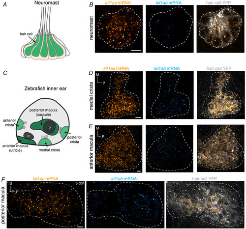
kif1aa mRNA is present in neuromast hair cells, while both kif1aa and kif1ab mRNAs are present in inner‐ear hair cells A,C, schematics showing a zebrafish neuromast and inner ear at 5 dpf. Within the inner ear, hair cells are present in three cristae and two maculae. Each macula is associated with an otolith (o). B, RNA‐FISH shows that at 5 dpf only kif1aa (orange) mRNAs are present in neuromast hair cells. D–F, in the inner ear, RNA‐FISH shows that at 5 dpf, both kif1aa (orange) and kif1ab (cyan) mRNAs are present in hair cells in cristae and maculae. The grey label is YFP, which is expressed specifically in hair cells by the transgenic line Tg(myo6b:Cr.ChR2‐EYFP). The YFP label was used to create the dashed line in B, D, E, F to outline the locations of hair cells within each sensory epithelium. Scale bars = 5 µm.
|