
Fig. 5
- ID
- ZDB-FIG-250430-169
- Publication
- Luo et al., 2025 - OPN3-mediated positive regulation of angiogenesis in HUVECs through VEGFR2 interaction
- Other Figures
- All Figure Page
- Back to All Figure Page
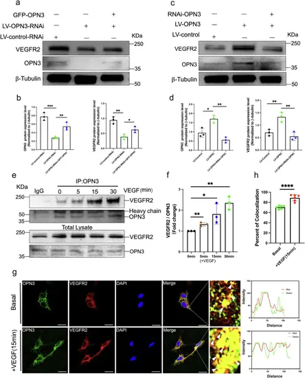
OPN3 and VEGFR2 are able to form a complex.a, b Western blot analysis was used to detect the protein levels of OPN3 and VEGFR2 in different cell groups with lentiviral knockdown of OPN3 or lentiviral knockdown of OPN3 combined with plasmid overexpression of OPN3 (GFP-OPN3), using β-tubulin as a loading control for normalization in the WB analysis. Relative protein levels were quantified using ImageJ software (n = 3 independent experiments, with each experimental group consisting of 6 dishes, derived from 3 different donors, with each donor providing 2 dishes of cells). Statistical analysis was performed using an unpaired t-test: ns (not significant), *p < 0.05, **p < 0.01, ***p < 0.001. c, d Western blot analysis was used to detect the protein levels of OPN3 and VEGFR2 in different cell groups with lentiviral overexpression of OPN3 or overexpression of OPN3 combined with siRNA-mediated knockdown of OPN3 (RNAi-OPN3), using β-tubulin as a loading control for normalization in the WB analysis. Relative protein levels were quantified using ImageJ software (n = 3 independent experiments, with each experimental group consisting of 6 dishes, derived from 3 different donors, with each donor providing 2 dishes of cells). Statistical analysis was performed using an unpaired t-test: *p < 0.05, **p < 0.01. e HUVECs were stimulated with VEGF (25 ng/mL), and cells were collected at different time points (5 min, 15 min, 30 min) to detect the interaction between OPN3 and VEGFR2 by Co-IP. Anti-OPN3 or anti-IgG (negative control) antibodies were used for immunoprecipitation (IP), followed by western blot analysis to detect OPN3 and VEGFR2 protein levels. Relative protein levels were quantified using ImageJ software. f Bar graph represents the averaged fold change of VEGFR2/OPN3 ratio over the basal ratio (n = 3 independent experiments, with each experimental group consisting of 12 dishes, derived from 6 different donors, with each donor providing 2 dishes of cells). Statistical analysis was performed using an unpaired t-test: *p < 0.05, **p < 0.01. g, h HUVECs were stimulated or not stimulated with VEGF for 15 min, followed by co-staining of OPN3 and VEGFR2. Images were captured using confocal microscopy. OPN3 was labeled in green, VEGFR2 in red, and DAPI in blue. The scale bar represents 25 μm. Yellow fluorescence (indicated by white arrows) in the merged images indicates their colocalization, which was analyzed by comparing the fluorescence intensity of each protein along the white line in the magnified images of the white box. A bar graph shows the percentage of colocalization (n = 6 dishes of cultured HUVECs from 3 different donors, with each donor replicated twice). Statistical analysis was performed using an unpaired t-test: ***p < 0.001. |