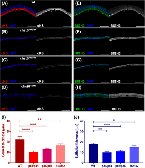
Corneal keratan sulfate (cKS) is reduced and BIGH3 expression is altered in the cornea of the adult mutant fish. Corneal sections stained with anti-cKS, anti-BIGH3, and DAPI are shown. (A) cKS (red) is detected in the corneal stroma of wt, but not detected in the mutant cornea as seen in images of (B) chst6pd4/pd4, (C) chst6pd5/pd5, (D) chst6f42/f42 mutant eye sections. Grayscale images of cKS signal are shown next to the overlay image; anti-cKS (red), DAPI (blue). (E) A strong stromal and weak epithelial BIGH3 (green) signal is detected in the cornea of the wt fish. BIGH3 is detected only in the epithelium of (F) chst6pd4/pd4 and (G) chst6pd5/pd5 mutants and (H) epithelium and stroma layers of the chst6f42/f42 mutant cornea. Scale bars: 80 μm. (I) Thickness of cornea (n = 5) and (J) corneal epithelium was measured from IF-stained cryosections represented in A–H (n = 5). Multiple t-test was used for the statistical analysis. Data are presented as the mean ± std. P < 0.05 (*), P < 0.01 (**), P < 0.001 (***), and P < 0.0001 (****) vs the wt group.
|
