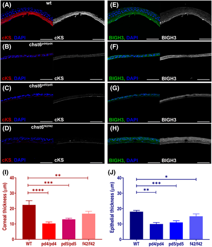
Fig. 8 Corneal keratan sulfate (cKS) is reduced and BIGH3 expression is altered in the cornea of the adult mutant fish. Corneal sections stained with anti-cKS, anti-BIGH3, and DAPI are shown. (A) cKS (red) is detected in the corneal stroma of wt, but not detected in the mutant cornea as seen in images of (B) chst6pd4/pd4, (C) chst6pd5/pd5, (D) chst6f42/f42 mutant eye sections. Grayscale images of cKS signal are shown next to the overlay image; anti-cKS (red), DAPI (blue). (E) A strong stromal and weak epithelial BIGH3 (green) signal is detected in the cornea of the wt fish. BIGH3 is detected only in the epithelium of (F) chst6pd4/pd4 and (G) chst6pd5/pd5 mutants and (H) epithelium and stroma layers of the chst6f42/f42 mutant cornea. Scale bars: 80 μm. (I) Thickness of cornea (n = 5) and (J) corneal epithelium was measured from IF-stained cryosections represented in A–H (n = 5). Multiple t-test was used for the statistical analysis. Data are presented as the mean ± std. P < 0.05 (*), P < 0.01 (**), P < 0.001 (***), and P < 0.0001 (****) vs the wt group.
Image
Figure Caption
Figure Data
Acknowledgments
This image is the copyrighted work of the attributed author or publisher, and
ZFIN has permission only to display this image to its users.
Additional permissions should be obtained from the applicable author or publisher of the image.
Full text @ FEBS J.

