|
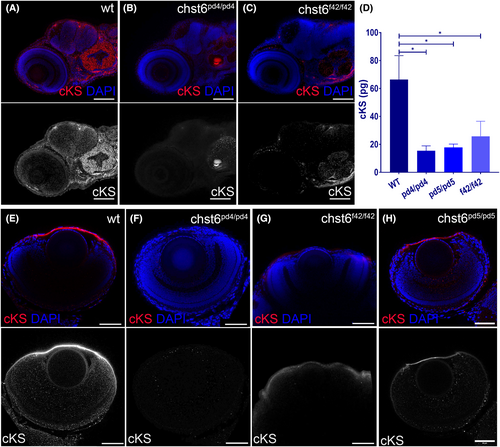
chst6 loss-of-function mutant phenotypes are rescued with wt mRNA. Loss of function was tested with cKS antibody using immunofluorescence and ELISA. (A) cKS epitopes are detected in the brain and otic vesicle of wt larvae at 4 dpf. The cKS signal is decreased in (B) chst6pd4/pd4 (C) chst6f42/f42 mutant larvae. (D) Total cKS in larval whole-body extracts were measured and displayed in the graph, and mean ± std values are wt: 66.36 ± 24.30 pg, chst6pd4/pd4: 15.46 ± 6 pg, chst6pd5/pd5: 17.83 ± 4.09 pg, chst6f42/f42: 25.81 ± 18.59 pg (n = 50, 3 replicates for each group, one-way ANOVA was used for statistical analysis). Corneal cKS signal is detected in wt (E). Lost in all mutants (F) chst6pd4/pd4, (G) chst6f42/f42 eye IF stainings are shown as examples. (H) cKS staining is rescued when wt mRNA is injected into 1-cell stage mutant embryos. P < 0.05 (*) Scale bars: 60 μm.
|