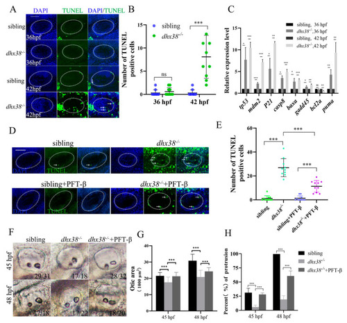
Apoptosis was increased in the inner ear epithelium cells of dhx38−/− zebrafish: (A) TUNEL staining in the inner ears of dhx38 mutants at 36 hpf and 42 hpf. The white arrows indicate the signal spots.The n = 10 for each panel. Scale bar: 50 µm. (B) Quantitative analysis of apoptotic cells of the inner ear (the white, dotted, oval area) region between sibling and dhx38−/− mutants. (C) The expression level of p53 pathway genes in sibling and dhx38−/− mutants at 36 hpf and 42 hpf using qPCR. (D) TUNEL staining indicated that cell apoptosis was inhibited in the dhx38 zebrafish mutant embryos by inhibiting p53. The white arrows indicate the signal spots. Scale bar: 50 µm. (E) Quantitative analysis of TUNEL-positive cells in the inner ear (the white, dotted, oval area) region of siblings, dhx38−/− homozygous embryos, and embryos with p53 inhibitor. (F) The inner ear morphology of different genotypes at 45 hpf and 48 hpf. (G) The statistical analysis of the otic lumen area at 45 hpf and 48 hpf. (H) The statistical proportion of protrusions at 45 hpf and 48 hpf. n = 30. The data are the mean ± SD. ns, p > 0.05; *, p < 0.05; **, p < 0.01; ***, p < 0.001.
|
