FIGURE

FIGURE 4

ID
ZDB-FIG-240222-86
Publication
Kobayashi-Sun et al., 2024 - Extremely low-frequency electromagnetic fields facilitate both osteoblast and osteoclast activity through Wnt/β-catenin signaling in the zebrafish scale
Other Figures
All Figure Page
Back to All Figure Page
FIGURE 4

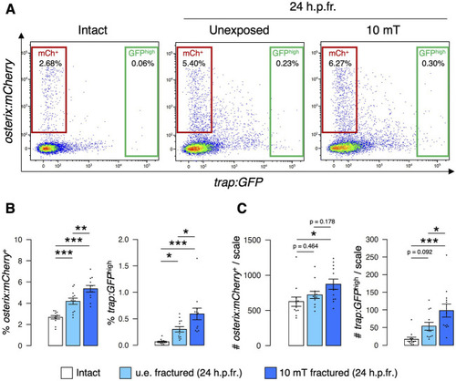
Both osteoblast and osteoclast numbers increase upon exposure to 10 mT of ELF-EMFs in the fractured scale. (A) Representative results of flow cytometric analysis of cells from intact scales (left panel), unexposed (u.e.) fractured scales (middle panel), or fractured scales exposed to 10 mT ELF-EMFs (right panel) at 24 h.p.fr. After fracture stimulation, zebrafish were placed in the ring-shaped tank and were unexposed or exposed to 10 mT ELF-EMFs for 4 h. Zebrafish scales were extracted at 24 h.p.fr., and cell samples were prepared for flow cytometric analysis. Red and green gates show trap:GFPosterix:mCherry+ (“mCh+”) and trap:GFPhigh (“GFPhigh”) cells, respectively. (B,C) Percentage (B) and absolute number of mCh+ and GFPhigh cells per scale (C) in intact scales, unexposed fractured scales (u.e. fractured), or fractured scales exposed to 10 mT ELF-EMFs (10 mT fractured). A total of 12 samples from 6 zebrafish were used in each condition. Asterisks indicate the p-value in one-way ANOVA with Dunnett’s test. Error bars, s.e.m.; *p < 0.05, **p < 0.01, ***p < 0.001.

Expression Data

Expression Detail
Antibody Labeling
Phenotype Data

Phenotype Detail
Acknowledgments
This image is the copyrighted work of the attributed author or publisher, and ZFIN has permission only to display this image to its users. Additional permissions should be obtained from the applicable author or publisher of the image. Full text @ Front Cell Dev Biol