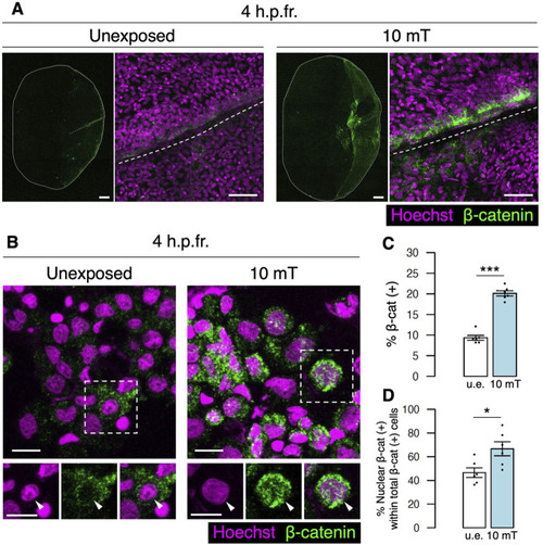
Expression of β-catenin is increased at the fracture site upon exposure to 10 mT of ELF-EMFs. (A) Expression of β-catenin in the fractured scale unexposed (u.e.) or exposed to 10 mT ELF-EMFs at 4 h.p.fr. After fracture stimulation, zebrafish were placed in the ring-shaped tank and were unexposed or exposed to 10 mT ELF-EMFs for 4 h. Zebrafish scales were then stained with rabbit anti-β-catenin antibody, followed by anti-rabbit IgG Alexa Fluor 647-conjugated secondary antibody and Hoechst 33342. White dotted lines and solid lines indicate the fracture site and contour of the zebrafish scale, respectively. Right panels show a high magnification view of the fracture site. Bars, 200 μm (left panels) and 40 μm (right panels). (B) Expression of β-catenin in cells from fractured scales unexposed or exposed to 10 mT ELF-EMFs at 4 h.p.fr. After fracture stimulation, zebrafish were placed in the ring-shaped tank and were unexposed or exposed to 10 mT ELF-EMFs for 4 h. Cells were then collected from zebrafish scales, smeared, and stained with rabbit anti-β-catenin antibody, followed by anti-rabbit IgG Alexa Fluor 647-conjugated secondary antibody and Hoechst 33342. Bottom panels show Hoechst (nuclei), β-catenin expression, and merged images of the dotted region. Arrowheads indicate the nucleus. Bars, 10 μm. (C,D) Percentage of β-catenin (+) cells within total cells and percentage of nuclear β-catenin (+) cells within total β-catenin (+) cells in the u.e. fractured scale or the fractured scale exposed to 10 mT ELF-EMFs at 4 h.p.fr. A total of 6 samples from 3 zebrafish were used in each condition. Asterisks indicate the p-value in unpaired two-tailed Student’s t-test. Error bars, s.e.m.; *p < 0.05.; ***p < 0.001.
|
