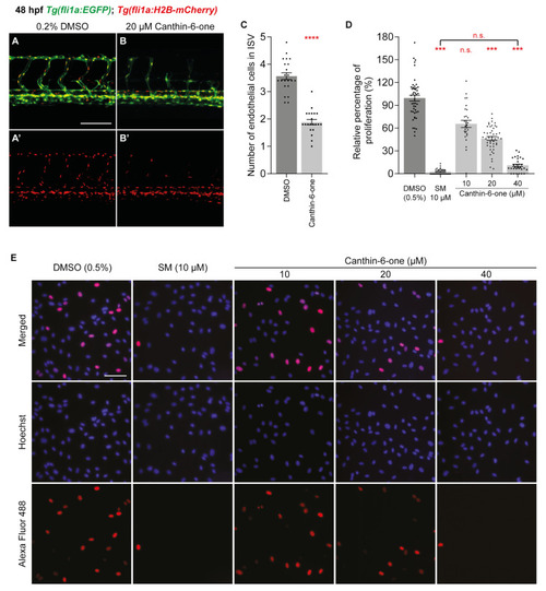
Canthin-6-one inhibits endothelial cell proliferation in zebrafish and HUVECs. (A,B’) Lateral confocal images of 48 hpf Tg(fli1a:EGFP);Tg(fli1a:H2B-mCherry) embryos treated with either 0.2% DMSO (A,A’) or 20 µM canthin-6-one (B,B’). Images (A’,B’) represent the fli1a:H2B-mCherry expression of images (A,B). (C) Quantification of intersegmental vessel (ISV) endothelial cell number in 48 hpf Tg(fli1a:EGFP);Tg(fli1a:H2B-mCherry) embryos treated with either 0.2% DMSO (n = 22 embryos) or 20 µM canthin-6-one (n = 21 embryos). Each data points represent an average of 5 ISVs from one embryo. (D) Quantification of relative percentage of proliferating HUVECs upon treatment with either 0.2% DMSO (n = 48 views), 10 µM sunitnib malate (SM, n = 30 views), or canthin-6-one at 10 µM (n = 28 views), 20 µM (n = 46 views), or 40 µM (n = 30 views). Results from at least 3 independent replicates. (E) Representative images of HUVECs treated with either 0.2% DMSO, 10 µM SM, or canthin-6-one at indicated concentrations. Cells were stained with Hoechst 33,342 (blue) and Alexa Fluor 488 (red) using the Click-iT assay. Blue indicates viable cells and red indicates proliferating cells. Statistical test: Mann-Whitney test for (C) and Kruskal-Wallis test for (D). p ≤ 0.0001 (****), p ≤ 0.001 (***), p > 0.05 (not significant, n.s.). Scale bars: 100 µm (A), 250 µm (E).
|
