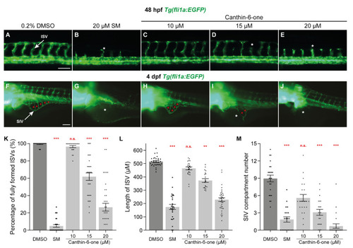
Canthin-6-one inhibits intersegmental vessel and sub-intestinal vessel development in zebrafish. (A–J) Lateral fluorescent images of 48 hpf (A–E) and 4 dpf (F–J) Tg(fli1a:EGFP) embryos treated with either 0.2% DMSO (A,F), 20 µM sunitinib malate (SM, B,G), or canthin-6-one at 10 µM (C,H), 15 µM (D,I), or 20 µM (E,J). White arrows indicate the intersegmental vessel (ISV, A) or the sub-intestinal vessel (SIV, F). Red asterisks indicate the vascular compartments within the SIV (F,H,I). White asterisks indicate the reduced/absence of ISVs (B,D,E) or SIV (G,I,J). (K) Quantification of the number of fully formed ISVs in 48 hpf Tg(fli1a:EGFP) embryos treated with either 0.2% DMSO (n = 30 embryos), 20 µM SM (n = 29 embryos), or canthin-6-one at 10 µM (n = 20 embryos), 15 µM (n = 29 embryos), or 20 µM (n = 37 embryos). (L) Quantification of the length of ISVs in 48 hpf Tg(fli1a:EGFP) embryos treated with either 0.2% DMSO (n = 32 embryos), 20 µM SM (n = 26 embryos), or canthin-6-one at 10 µM (n = 25 embryos), 15 µM (n = 20 embryos), or 20 µM (n = 37 embryos). (M) Quantification of the number of SIV compartments in 4 dpf Tg(fli1a:EGFP) larvae treated with either 0.2% DMSO (n = 23 larvae), 20 µM SM (n = 20 larvae), or canthin-6-one at 10 µM (n = 22 embryos), 15 µM (n = 21 embryos), or 20 µM (n = 24 embryos). Statistical test: Kruskal-Wallis test was conducted for graphs (K–M). p ≤ 0.001 (***), p ≤ 0.01 (**), p > 0.05 (not significant, n.s.). Scale bars: 100 µm.
|
