
Figure 1—figure supplement 6
- ID
- ZDB-FIG-231116-87
- Publication
- Shrestha et al., 2023 - The myocardium utilizes a platelet-derived growth factor receptor alpha (Pdgfra) - phosphoinositide 3-kinase (PI3K) signaling cascade to steer towards the midline during zebrafish heart tube formation
- Other Figures
-
- Figure 1
- Figure 1—figure supplement 1
- Figure 1—figure supplement 2
- Figure 1—figure supplement 3
- Figure 1—figure supplement 4
- Figure 1—figure supplement 5
- Figure 1—figure supplement 6
- Figure 2
- Figure 2—figure supplement 1
- Figure 2—figure supplement 2
- Figure 3
- Figure 3—figure supplement 1
- Figure 3—figure supplement 2.
- Figure 4
- Figure 4—figure supplement 1
- Figure 5
- All Figure Page
- Back to All Figure Page
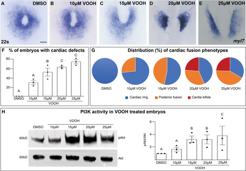
Inhibition of Pten activity with VO-OHpic increases pAkt and causes cardiac fusion defects. (A–G) Dorsal views of the myocardium labeled with myl7 at 22s in embryos incubated with increasing concentrations of the Pten inhibitor VO-OHpic (VO-OH) from bud stage to 22s. Graphs depicting the average % of embryos displaying cardiac fusion defects (F) and the distribution of cardiac fusion phenotypes (G). Blue – cardiac ring/normal; orange – fusion only at posterior end/mild phenotype, red – cardia bifida/severe phenotype. (H) Representative immunoblot and graph of ratiometric analysis of pAKT to AKT protein levels indicates increasing pAKT levels with increasing concentrations of the Pten inhibitor VO-OH. Three separate incubations per concentration (dots in F, H). n = 15 embryos per incubation per concentration (A–G). Letter change indicates p < 0.05, one-way analysis of variance (ANOVA). Raw data and full p-values included in the source file. |