
Figure 1—figure supplement 3
- ID
- ZDB-FIG-231116-84
- Publication
- Shrestha et al., 2023 - The myocardium utilizes a platelet-derived growth factor receptor alpha (Pdgfra) - phosphoinositide 3-kinase (PI3K) signaling cascade to steer towards the midline during zebrafish heart tube formation
- Other Figures
-
- Figure 1
- Figure 1—figure supplement 1
- Figure 1—figure supplement 2
- Figure 1—figure supplement 3
- Figure 1—figure supplement 4
- Figure 1—figure supplement 5
- Figure 1—figure supplement 6
- Figure 2
- Figure 2—figure supplement 1
- Figure 2—figure supplement 2
- Figure 3
- Figure 3—figure supplement 1
- Figure 3—figure supplement 2.
- Figure 4
- Figure 4—figure supplement 1
- Figure 5
- All Figure Page
- Back to All Figure Page
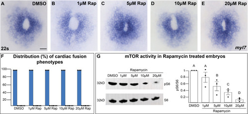
Inhibition of mTOR activity does not affect cardiac fusion. (A–E) Myocardium visualized with myl7 expression at 22s in embryos treated at bud stage with increasing concentrations of rapamycin (Rap), an inhibitor of mTOR activity. (F) Bar graph displays the distribution of cardiac phenotypes at each rapamycin concentration from three replicates. Total number of embryos analyzed n = 45, 46, 45, 45, 44, respectively. Blue bar = cardiac ring/normal; scale bar = 40 μm. All embryos display cardiac rings, indicating normal cardiac fusion. (G) Representative immunoblot and ratiometric analysis of phosphorylated ribosomal protein S6 (pS6) – a read-out of mTOR activity, to S6 levels reveals that mTOR activity decreases with increasing concentrations of rapamycin. Letter change indicates p < 0.05, one-way analysis of variance (ANOVA). Raw data with full p-values included in the source file. |