FIGURE

Figure 1—figure supplement 2

ID
ZDB-FIG-231116-83
Publication
Shrestha et al., 2023 - The myocardium utilizes a platelet-derived growth factor receptor alpha (Pdgfra) - phosphoinositide 3-kinase (PI3K) signaling cascade to steer towards the midline during zebrafish heart tube formation
Other Figures
All Figure Page
Back to All Figure Page
Figure 1—figure supplement 2

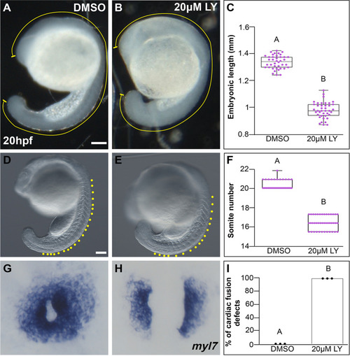
LY incubation results in trunk extension and somite formation delays.

Lateral brightfield views of 20 hours post-fertilization (hpf) embryos treated with DMSO (A, D) or 20 μM LY (B, E) at bud stage. (C, F) Box-whisker plot depicting the median embryonic length (yellow curved line in A, B) or somite number (yellow dots in D, E) at 20 hpf. Total number of embryos (n) from >3 separate incubations = 40 (DMSO), 40 (20 μM LY) for (C), and 39 (DMSO), 42 (20 μM LY) for (F). Dots = measurements from individual embryos. Two-sample t-test; p-value = 4.527 × 10⁻⁴ and 7.624 × 10⁻⁵, respectively. (G–H) Dorsal views, anterior to the top, of the myocardium labeled with myl7 at 20 hpf. Embryos treated with DMSO at bud stage show cardiac rings (G) whereas those treated with 20 μM LY show cardia bifida at 20 hpf (H). (I) Graph depicts the average percentage of cardiac fusion defects in embryos treated with DMSO or 20 μM LY. The total number of embryos examined from three separate incubations (n) = 45 (DMSO), 45 (20 μM LY). Two-sample t-test; p-value = 4.56 × 10⁻⁵. Dots indicate the percent of embryos with cardiac fusion defects per incubation. Letter changes (C, F, I) indicate p-values <0.05. Raw data included in the source file.

Expression Data

Expression Detail
Antibody Labeling
Phenotype Data

Phenotype Detail
Acknowledgments
This image is the copyrighted work of the attributed author or publisher, and ZFIN has permission only to display this image to its users. Additional permissions should be obtained from the applicable author or publisher of the image. Full text @ Elife