FIGURE

Figure 1—figure supplement 5

ID
ZDB-FIG-231116-86
Publication
Shrestha et al., 2023 - The myocardium utilizes a platelet-derived growth factor receptor alpha (Pdgfra) - phosphoinositide 3-kinase (PI3K) signaling cascade to steer towards the midline during zebrafish heart tube formation
Other Figures
All Figure Page
Back to All Figure Page
Figure 1—figure supplement 5

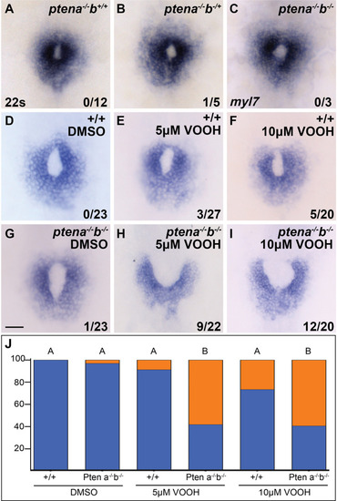
Loss of Pten, an antagonist of phosphoinositide 3-kinase (PI3K) activity, causes cardiac fusion defects.

(A–I) Dorsal views, anterior to the top, of the myocardium labeled with myl7 at 22s. Neither ptena−/− homozygous mutants (A), ptena−/− homozygous; ptenb−/+ heterozygous (B), nor ptena−/−, ptenb−/− double homozygous mutants (C), display cardiac fusion defects. However, maternal contribution of Pten has been reported to persist during development. Adding low concentrations of the Pten inhibitor VO-OHpic (VO-OH – 5, 10 μM) at bud stage to ptena−/−, ptenb−/− double homozygous mutants to inhibit maternal Pten activity did cause a significant increase in cardiac fusion defects (H, I), compared to DMSO (D, G) and low concentrations of VO-OH only (E, F). The number of embryos with cardiac fusion defects and total number analyzed are indicated. (J) Bar graph depicts the distribution of cardiac fusion defects (% of embryos analyzed) in wild-type or ptena−/−, ptenb−/− double homozygous mutants, treated with DMSO, or 5, 10 μM VO-OH. Blue – cardiac ring/normal; orange – fusion only at posterior end. Fisher’s exact test, letter change indicates p < 0.05. Scale bar, 40 μm. Raw data including quantification of all genotypes and full p-values included in the source file

Expression Data

Expression Detail
Antibody Labeling
Phenotype Data

Phenotype Detail
Acknowledgments
This image is the copyrighted work of the attributed author or publisher, and ZFIN has permission only to display this image to its users. Additional permissions should be obtained from the applicable author or publisher of the image. Full text @ Elife